Siemens ACUSON X700
Siemens Healthineers · ACUSON series
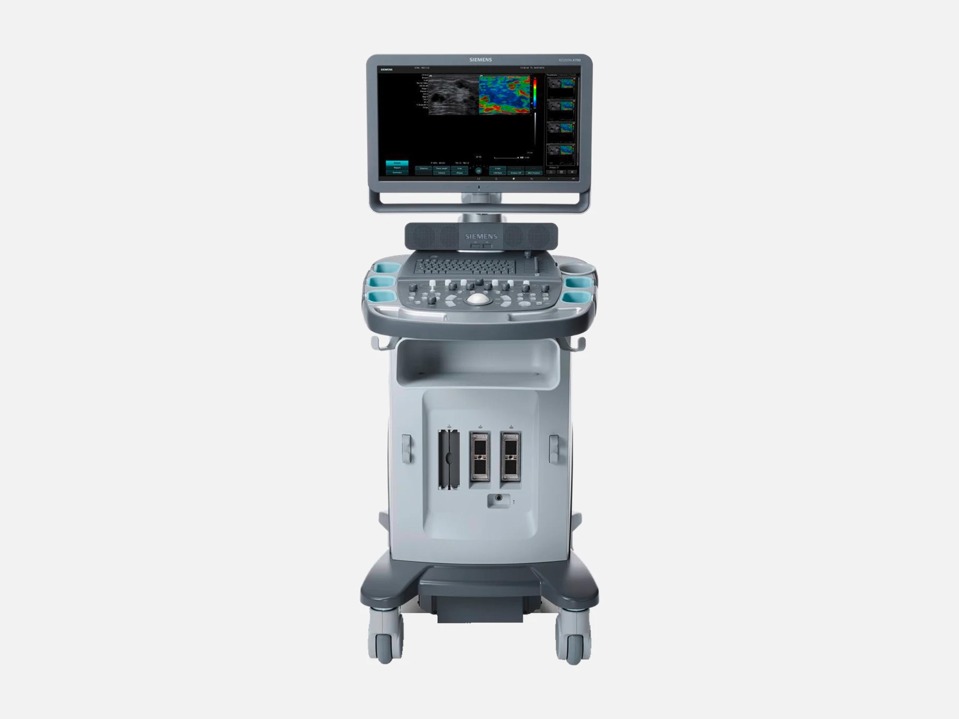
The Siemens ACUSON X700 is a cart-based ultrasound system for shared-service imaging and cardiology, launched in 2012 as the top model of the ACUSON X Family. It uses an imaging engine from the ACUSON S Family, accepts S Family and X Family transducers and runs TEE and intracardiac probes. It shares a 2014 FDA clearance with the ACUSON X600, which lacks its eSie Touch elasticity and syngo Auto Follicle options.
- Released
- 2012launched by Siemens
- Probe ports
- 3all active
- Display
- 20-inch1600 x 900 widescreen
- Weight
- 102 kg225 lb, approximately
Compatible probes
Linear
- VF10-5 · 5–10 MHz
- VF12-4 · 4–12 MHz Long linear
- VF16-5 · 5–15 MHz
Convex & microconvex
- 4C1 · 1–5 MHz
- 6C2 · 2–6 MHz
- C6F2 · 1.5–5.5 MHz fourSight 4D
- C8F3 · 2–7 MHz fourSight 4D
- C7F2 Mechanical 3D/4D
- EV9F4 Mechanical 3D/4D
- C8-5
- MC9-4 · 4–9 MHz Wide field of view
- EC9-4w · 4–9 MHz
Phased
- 4V1c · 1–4 MHz
- P8-4 · 2.7–8 MHz
Endocavity
- EV9F3 · 3–9 MHz Endovaginal, fourSight 4D
Specialty
- V5Ms · 3–7 MHz Transesophageal
- AcuNav 8F · 5–10 MHz Intracardiac catheter, 8 French
- AcuNav 10F · 5–10 MHz Intracardiac catheter, 10 French
- SoundStar 10F Intracardiac catheter
- SoundStar eco 10F Intracardiac catheter
- SoundStar eco 8F Intracardiac catheter
- CW2 CW Doppler, 2.0 MHz
- CW5 CW Doppler, 5.0 MHz
Specifications
Display
- Display
20-inch widescreen LED/LCD flat panel, IPS, 1600 x 900 pixels, 178° angle of view20-inch widescreen flat panel display (LED/LCD), color, high resolution, progressive scan (non-interlaced), In Plane Switching (IPS) technology; resolution 1600 x 900 pixels; angle of view 178°Show allShow less
- Monitor arm
Articulating arm: rotation ±90°, panel ±80°, tilt 15° up / 90° down, swivel ±80°Multi-directional 180° articulating monitor arm with integrated handle: arm rotation -90° to +90°, flat panel rotation -80° to +80°, up 125 mm, pull 250 mm; monitor tilt 15° up and 90° down, swivel ±80°Show allShow less
- Display formats
Full screen, Split, Quad, Dual, Dual Select, Dual from Freeze, 4B-mode, Curved VectorFull screen, Split, Quad, Dual, Dual Select and Dual from Freeze formats; 4B-mode (4 static B-mode images); Curved Vector formatShow allShow less
Physical
- Weight
- Approximately 102 kg (225 lb); 98 kg (216 lb) without OEM devices
- Dimensions
Height 138.6–166.3 cm, width 55.6 cm, depth 90.5 cm (97.9 cm with rear handle)Height 138.6–166.3 cm (54.6–65.5 in); width 55.6 cm (21.9 in); depth 90.5 cm (35.6 in) without rear handle, 97.9 cm (38.5 in) with rear handleShow allShow less
- Wheels
- Four 125 mm casters; front pair bi-brake (direction and total lock), rear total lock
- Probe ports
3 active ports for phased, curved and linear arrays; 512 MP and DL (260) connectors3 active transducer ports supporting high density phased array, curved array and linear array transducers; electronic transducer selection; 512 micro-pinless (MP) and DL (260) type connectorsShow allShow less
- Control panel
Swivels ±90° and rises 10 cm; backlit keys, customizable soft keys, QWERTY keyboardHeight-adjustable, swivel control panel with lock lever; Home Base design; left/right swivel articulation ±90°, vertical adjustment 10 cm; backlit key illumination; customizable soft keys; full-size QWERTY keyboardShow allShow less
- Transducer holders
6 configurable holders, gel bottle storage, endocavity holder; removable for cleaning6 configurable transducer holders with gel bottle storage; special holder for the endocavity transducer; holders removable for cleaningShow allShow less
Power & battery
- Power
100–120/200–240 VAC, 50/60 Hz; isolation transformer; max 600 VA with OEM devicesOne mainframe configuration for 100/115 V and 230 V environments; power connections 100–120/200–240 VAC, 50/60 Hz; built-in AC isolation transformer; power consumption maximum 600 VA with OEM devicesShow allShow less
- QuikStart (option)
- Standby mode: powers down in 3 sec, boots fully in less than 12 sec
Connectivity & options
- DICOM
DICOM 3.0 Store, Print, Worklist, MPPS, storage commitment, query/retrieve, SRDICOM 3.0: DICOM Storage Class User and DICOM Print Class User; DICOM Print/Store, Modality Worklist, MPPS, storage commitment and query/retrieve; DICOM structured reporting, including OB/GYN and VascularShow allShow less
- DICOM Structured Reporting (option)
- For OB/GYN and Vascular applications
- Network
- One RJ45 Ethernet connector, 10/100 BaseT
- Wireless Data Transfer (option)
USB dongle to facility LAN; IEEE 802.11n/g/b/a; WEP, WPA and WPA2 securityUSB dongle connecting to the facility LAN; IEEE 802.11n/g/b/a; WEP, WPA, WPA2 personal, WPA and WPA2 Enterprise; supports DICOM services, including wireless PACS, and Siemens Remote ServiceShow allShow less
- USB ports
- 6 USB 2.0 ports: 2 front, 4 rear
- Video and audio
- VGA out, DVI out, DVI in/out, external audio out
- Storage
- 500 GB hard drive, 300 GB for patient data; up to 300,000 images with compression
- Removable media
- Built-in DVD multi-drive (DVD-R/RW, CD-R/RW); export to USB flash drive
- Export formats
- RTF, PDF, TIFF, AVI, JPG and DICOM
- Documentation devices
Up to 2 built into the cart: B/W printer and color printer or DVR, run from the panelUp to 2 documentation devices (B/W printer and color printer or DVR) integrated into the cart and controlled from the control panel; supported: P95DW and UP-D897 B/W printers, CP30DW and UP-D25MD color printers, UR-50BDS DVRShow allShow less
- Barcode reader (option)
- 1D and 2D patient barcodes, up to 3 (patient, physician, sonographer); USB
- Virus protection (option)
- Embedded virus protection (Ultrasound System Security)
- Integrated gel warmer (option)
- Low 31 °C, Medium 34 °C, High 37 °C; temperature precision ±1°
Imaging & software
- Imaging modes
B, M, anatomical M, color M, color, power and PW/CW Doppler, tissue Doppler, THI, 3D/4D2D (fundamental 2D; phased, filtered and alternating THI); M-mode, color M-mode and anatomical M-mode; velocity-based color Doppler, power Doppler, directional power Doppler and color Doppler tissue imaging; pulsed wave and continuous wave Doppler, spectral Doppler tissue imaging, duplex and triplex modes; amplitude Doppler; combined modes; harmonic imaging with 3D and 4D imagingShow allShow less
- Revision and software
- Release 2.0; software versions 1.0/1.1 and VB20
- System architecture
1.0–17.0 MHz; up to 512 lines; up to 23,360 processing channels; > 205 dB dynamic rangeSystem imaging frequency 1.0–17.0 MHz; 2D-mode line density up to 512 lines; up to 23,360 processing channels; total system dynamic range > 205 dBShow allShow less
- Transducer frequency range
- 1.3–16.0 MHz
- 2D frame rate
- Up to 498 fps (transducer dependent)
- Display depth
- 1 cm to 30 cm; up to 4 transmit focal zones
- Cine memory
- Up to 2729 frames (standard)
- Clip capture
- Retrospective 1, 2 or 4 seconds; prospective 1 to 120 seconds
- Built-in imaging technologies
Native THI, DTO, Dynamic TCE, SieScape, Advanced SieClear and SieClear compoundingNative tissue harmonic imaging, DTO Dynamic Tissue Optimization, Dynamic TCE tissue contrast enhancement, SieScape panoramic imaging, Advanced SieClear spatial compounding and SieClear compoundingShow allShow less
- Advanced SieClear
- Spatial compounding, up to 7 steering angles on linear and 5 on curved arrays
- Dynamic TCE
- Speckle-reduction post-processing at Low, Medium or High
- SieScape
- Grayscale panoramic images up to 60 cm long, curvature up to 360°
- TGO
- One-button optimization of image brightness and gain for the tissue type
- MultiHertz
Up to 7 transmit frequencies: 4 in 2D and M-mode, 4 in THI, 2 in color, power and PWUp to 7 user-selectable transmit frequencies: 2D and M-mode up to 4 fundamental, THI up to 4, color, power and PW Doppler up to 2Show allShow less
- QuickSet
- Up to 32 user presets of imaging parameters, annotation, text and measurements
- Clarify VE
- Vascular enhancement on all curved and linear transducers
- 3D and 4D
3-Scape real-time freehand 3D (4C1, 6C2); fourSight 4D (C6F2, C8F3, EV9F3)3-Scape real-time 3D imaging, freehand acquisition, on the 4C1 and 6C2 transducers; fourSight 4D transducer technology (C6F2, C8F3 and EV9F3) and Advanced fourSight technologyShow allShow less
- Further imaging tools
- Dual HD Zoom, Ensemble Tissue Harmonic Imaging, DTI Doppler tissue imaging
- Doppler automation
- AutoColor flow state optimization; auto-trace Doppler measurements
- Cardiac tools
- syngo Auto Left Heart, Axius edge-assisted ejection fraction, stress echo
- syngo Velocity Vector Imaging (option)
- Myocardial motion and mechanics from a 2D image: rotation, strain and strain rate
- syngo Arterial Health Package
- Carotid intima-media thickness (CIMT) measurement
- syngo Auto OB and Auto Follicle (options)
- Auto OB: BPD, HC, AC, FL and HL; Auto Follicle: multiple follicles
- eSie Touch elasticity imaging (option)
- Real-time elastogram of relative tissue stiffness, from gentle compression in B-mode
- Cadence contrast agent imaging (option)
- Optimized for the 4C1 transducer for abdominal applications
- DIMAQ-IP
- Integrated workstation: on-board acquisition, storage and review of studies
Environment
- Atmospheric pressure
- 700–1060 hPa
- Heat output
- Maximum 2150 BTU/hr
- Noise level
- Average 46–47 dB
Compliance
- FDA 510(k)
K141846 (2014-08-07), K123001 (2012-10-26)K141846 (decided 2014-08-07; ACUSON X700 and ACUSON X600); K123001 (decided 2012-10-26). Class II, product code IYN, 21 CFR 892.1550.Show allShow less
- Quality and safety standards
21 CFR Part 820, ISO 9001, ISO 13485; UL 60601-1, CSA C22.2 No. 601-1, EN/IEC 60601-1Quality: QSR 21 CFR Part 820, ISO 9001, ISO 13485. Design standards include UL 60601-1, CSA C22.2 No. 601-1 and EN/IEC 60601-1.Show allShow less
What to check when buying a used Siemens ACUSON X700
Inspect the console for fluid and chemical damage
Fluid that seeps into the electrical circuitry can cause excessive electrical leakage or system failure, and disinfectants not specified for the system can damage its surfaces and create electrical hazards.
How: Look along the control panel seams, the keyboard, the trackball area, the DVD slot and the outer surfaces for signs of fluid ingress or chemical damage.
Check the OB/GYN report recall, Z-1504-2014
On software 1.0.00, 1.0.01, 1.0.02, 1.1.00 and 1.1.02, using fourSight 4D, 3-Scape 3D or anatomical M-mode in an obstetric or gynecology study could put the previous patient's data into the report.
How: Read the installed software version, check the correction records for the recall, and confirm the software upgrade was applied.
Inspect the V5Ms articulating section and test leakage
Recall Z-2571-2014 concerned deterioration of the material covering the V5Ms articulating section, a risk avoided only when the leakage test in the user manual is performed.
How: If a V5Ms comes with the unit, check that covering for damage or wear and have the leakage test run, and reject a probe that shows either fault or fails the test.
Confirm the V5Ms lock-up fix, recall Z-1572-2016
On software 1.0.04 or 1.1.04, a communication error with the V5Ms rotation function could show an IMG_15 error and lock up the system until a reboot.
How: Read the installed software version on any unit used with a V5Ms, and confirm the correction to software 1.0.05 under update program US020/15/S was applied.
Common questions about the Siemens ACUSON X700
Is the Siemens ACUSON X700 FDA-cleared?
Yes. The ACUSON X700 is FDA-cleared under 510(k) K123001 (decided October 26, 2012) and K141846 (decided August 7, 2014), and it shares K141846 with the ACUSON X600. It is a Class II device under 21 CFR 892.1550, product code IYN.
Is the Siemens ACUSON X700 discontinued?
Yes. The ACUSON X700 is among Siemens' ultrasound legacy systems and is no longer commercially available. Its commercial distribution end date in the FDA's device database is December 31, 2020.
What is the difference between the Siemens ACUSON X700 and X600?
The X700 was launched in 2012 as the top model of the ACUSON X Family. It shares the 2014 FDA clearance K141846 with the ACUSON X600, and of the two, only the X700 offers syngo Auto Follicle measurements and eSie Touch elasticity imaging.
In that clearance, the indications for the CH5-2 and C6-2 transducers name the X600, not the X700.
Can the Siemens ACUSON X700 use ACUSON S Family transducers?
Yes. The X700 accepts transducers from both the ACUSON X Family and the ACUSON S Family, and its imaging engine was migrated from the S Family.
Its 3 active transducer ports take phased, curved and linear array transducers, with 512 micro-pinless (MP) and DL (260) connectors.