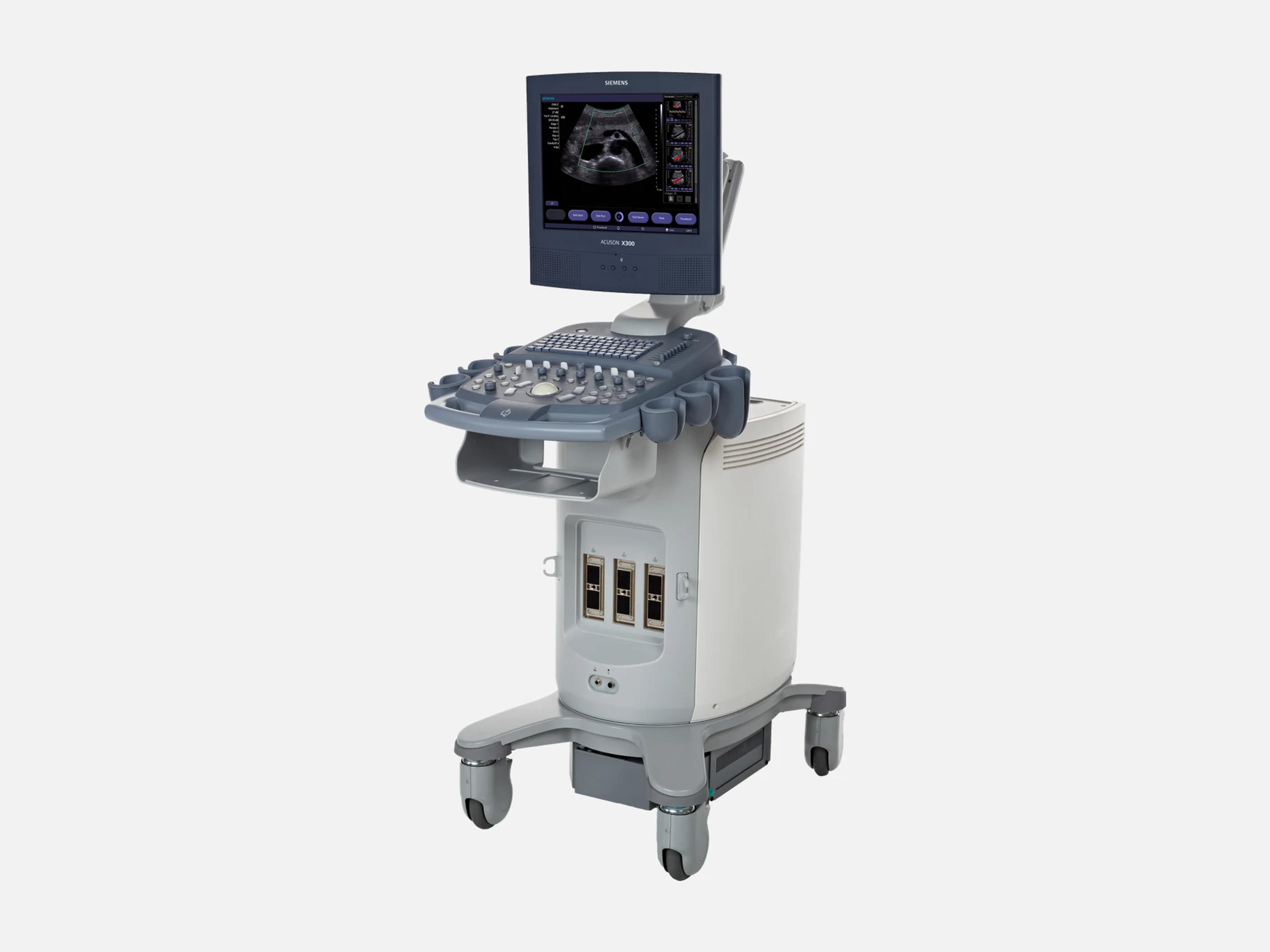
The Siemens ACUSON X300 PE is the premium edition of the ACUSON X300, a cardiology platform that also serves general imaging, OB/GYN, urology and emergency medicine. Where the ACUSON X300 has a 15-inch display and MultiBeam, the PE has an 18-inch display, Quad-Beam formation and a Plus Option that doubles channel count. It runs TEE and 3D TEE, AcuNav intracardiac imaging and Velocity Vector Imaging strain.
- Released
- 2008first FDA clearance
- Display
- 18-inchflat panel, articulating arm
Compatible probes
Linear
- VF8-3
- VF10-5
- VF13-5
- VF13-5SP
Convex & microconvex
- C6-2
- CH5-2
- C8-5
- C7F2
Phased
- P5-1 Single crystal
- P4-2
- P8-4
- P9-4
Endocavity
- EC9-4
- EV9-4
- EV9F4
- BP9-4 Live bi-plane prostate imaging
Specialty
- V5Ms Transesophageal
- CW2 Continuous-wave Doppler, 2.0 MHz
- CW5 Continuous-wave Doppler, 5.0 MHz
- AcuNav 8F Intracardiac catheter
- AcuNav 10F Intracardiac catheter
- SoundStar Intracardiac catheter for CartoSound
- P4-2 Litho LITHOSKOP lithotripter localization
Specifications
Display
- Display
18-inch high-resolution flat-panel display, fully adjustable, on an articulating arm18-inch high-resolution flat-panel display, fully adjustable, with an integrated handle on the monitor; mounted on an articulating armShow allShow less
- Display formats
- Split/Zoom view (reference and zoomed image together); Dual Screen Imaging
Physical
- Control panel
Height-adjustable control panel; customizable panel, menus, measurements and reportsHeight-adjustable control panel; user-customizable control panel, menus, measurements and reports; console built around a home-base design; ErgoDynamic imaging system designShow allShow less
Power & battery
- Battery
- Battery operation, optional
- QuikStart
- Rapid boot
Connectivity & options
- Archiving
- CD/DVD, USB or DICOM-enabled networks
- DICOM
- Full DICOM connectivity, including DICOM SR, and DICOM worklist
- File format
- Universal file format, online and offline
- Data entry
- Patient search function and barcode reader
- CartoSound Communication
Ethernet link to CartoSound; Carto 3 System integration; SoundStar 3D catheter imagingEthernet communication between the ACUSON X300 PE and the CartoSound system; integration with Carto 3 System technology and SoundStar 3D catheter imagingShow allShow less
- Plus Option
Hardware option doubling the system channel count, for high-density transducersBeamforming hardware option that doubles the system channel count; for high-density transducers on the ACUSON X300 PEShow allShow less
- LITHOSKOP interface
LITHOSKOP interface SW and P4-2 Litho transducer for in-line ultrasound localizationP4-2 Litho transducer and LITHOSKOP interface SW: options and transducer for in-line ultrasound localization with the Siemens LITHOSKOP lithotripterShow allShow less
Imaging & software
- Imaging modes
B-mode, color Doppler, color and anatomical M-mode, steerable CW, spectral and color DTIB-mode, color Doppler and mixed modes; color M-mode; anatomical (angle-corrected) M-mode, available during live, cineloop and DIMAQ image review; steerable continuous wave Doppler; spectral and color DTI Doppler tissue imagingShow allShow less
- Further modes
Ensemble tissue harmonic imaging; B&W and Power SieScape panoramic; Tissue EqualizationEnsemble tissue harmonic imaging, 3D imaging, B&W SieScape panoramic imaging, Power SieScape panoramic imaging, contrast agent imaging, SieClear multi-view spatial compounding, Tissue Equalization Technology, intracardiac imagingShow allShow less
- Quad-Beam formation technology
- Parallel signal processing for frame rate in B-mode, color Doppler and mixed modes
- Hanafy lens and SynAps technology
Abdominal imaging to a depth of 30 cm with Hanafy lens transducer technologyHanafy lens transducer technology and SynAps synthetic aperture technology; abdominal imaging to a depth of 30 cm with Hanafy lens transducer technologyShow allShow less
- Dynamic TCE
- Real-time processing that enhances tissue contrast resolution and reduces speckle
- TGO
- Tissue Grayscale Optimization: one-button image optimization
- Clarify VE
- Vascular enhancement that reduces noise and artifacts within the vessel
- SieClear
- Multi-view spatial compounding
- Contrast imaging
Cadence contrast agent imaging; one-button LVO contrast; low-MI THI setting for LVOCadence contrast agent imaging technology; one-button LVO contrast; a low Mechanical Index THI setting for LVO contrast imaging. At publication, ultrasound contrast agents were cleared in the US only for LVO.Show allShow less
- 3D/4D imaging
Advanced fourSight 3D/4D; fourSight 4D transducers; TEE and 3D TEE (fourSight TEE View)Advanced fourSight technology (3D/4D); fourSight 4D transducer technology; TEE and 3D TEE using syngo fourSight TEE ViewShow allShow less
- syngo fourSight TEE View
- 3D display of cardiac structure and function
- syngo Velocity Vector Imaging (VVI)
2D tracking for rotation, strain and strain rate (longitudinal, circumferential, radial)2D tracking algorithm for rotation, strain and strain rate calculations (longitudinal, circumferential and radial) of myocardial mechanicsShow allShow less
- syngo Auto Left Heart (Auto LH)
- Automatically calculates left atrial and left ventricular volumes and ejection fractions
- syngo Mitral Valve Assessment (MVA)
- 3D anatomical mapping of mitral valve structure, spatial relationships and blood flow
- syngo Arterial Health Package (AHP)
Semi-automated CIMT measurement, for use per the ASE carotid consensus statementSemi-automated carotid intima-media thickness (CIMT) measurement, for use per the ASE Consensus Statement on carotid ultrasoundShow allShow less
- syngo Auto OB
- Knowledge-based OB measurements that recognize features and patterns in the image
- Intracardiac echocardiography (ICE)
- ICE imaging with an 8F or 10F ACUSON AcuNav intracardiac transducer
- Live dual Bi-Plane
Simultaneous bi-plane prostate imaging with the BP9-4; dual-needle-path biopsy modeLive simultaneous bi-plane prostate imaging with the BP9-4 transducer; biopsy mode with dual needle pathsShow allShow less
- Reporting packages
Cardiac package; physician summary report; OB/GYN, Vascular, Endocrinology and moreCardiac imaging package; customizable physician summary report package with on-system report generation; specialized reporting packages for Endocrinology, Follicle Assessment, OB/GYN, Vascular, Cardiology, Emergency Medicine and moreShow allShow less
- US syngo Workplace
- syngo modules for analyzing scanned images on a personal computer
- Revision and software
- Release 7.0; software versions 3.0.00, 3.0.02, 5.0.00 and 5.0.01
Compliance
- FDA 510(k)
- K080760 (April 25, 2008); K121699 (June 22, 2012); Class II
What to check when buying a used Siemens ACUSON X300 PE
Watch power-up for smoke or a burning smell
Units have given off smoke and a burning smell at power-up from standby, traced to a faulty AC power assembly, and one tripped the facility's fuse.
How: Start the system from fully off and again from standby, and treat any odor or smoke as an AC power assembly fault to fix before purchase.
Confirm the cardiac PGmean recall correction is installed
Recall Z-1838-2011 covered every software version with cardiovascular options installed, because the cardiac package underestimated the mean pressure gradient traced from a stenotic Doppler waveform.
How: On a unit with cardiovascular options, ask for the service record showing this recall's correction was applied.
Confirm the V5Ms shutdown software update
On software 5.0.00 and 5.0.01, the V5Ms transesophageal transducer with the Plus option caused an electrical overload fault that shut the system down, the subject of recall Z-2061-2011.
How: On a unit with the V5Ms and the Plus option, check the service record for the field software update, then image in CW Doppler and confirm the system stays on.
Check vascular reports for left-right data swaps
On software 3.0.00 and 3.0.02, a user-defined formula in a vascular exam put data on the wrong side of the summary report, the subject of recall Z-2147-2011.
How: Read the installed software version, ask for the mandatory update's record, and run a vascular exam with a user-defined formula to confirm each side reports correctly.
Common questions about the Siemens ACUSON X300 PE
Is the Siemens ACUSON X300 PE FDA-cleared?
Yes. It is FDA-cleared through 510(k) K080760, decided April 25, 2008, and K121699, decided June 22, 2012, both of which name the ACUSON X300 premium edition among their device names. It is a Class II device.
Is the Siemens ACUSON X300 PE discontinued?
Yes. Its US commercial distribution ended on December 31, 2018.
Does every ACUSON X300 PE support Velocity Vector Imaging?
No. On the X300 line, syngo Velocity Vector Imaging is limited to the premium edition with a Cardiovascular or Advanced Cardiovascular package, and further technical prerequisites can apply, so confirm it is installed on the unit itself.
Is there an OB/GYN version of the ACUSON X300 PE?
Yes. Siemens introduced the ACUSON X300 PE Women's Imaging configuration on July 1, 2009, with 3D/4D imaging and syngo Auto OB automated fetal biometry.