Siemens ACUSON X300
Siemens Healthineers · ACUSON series
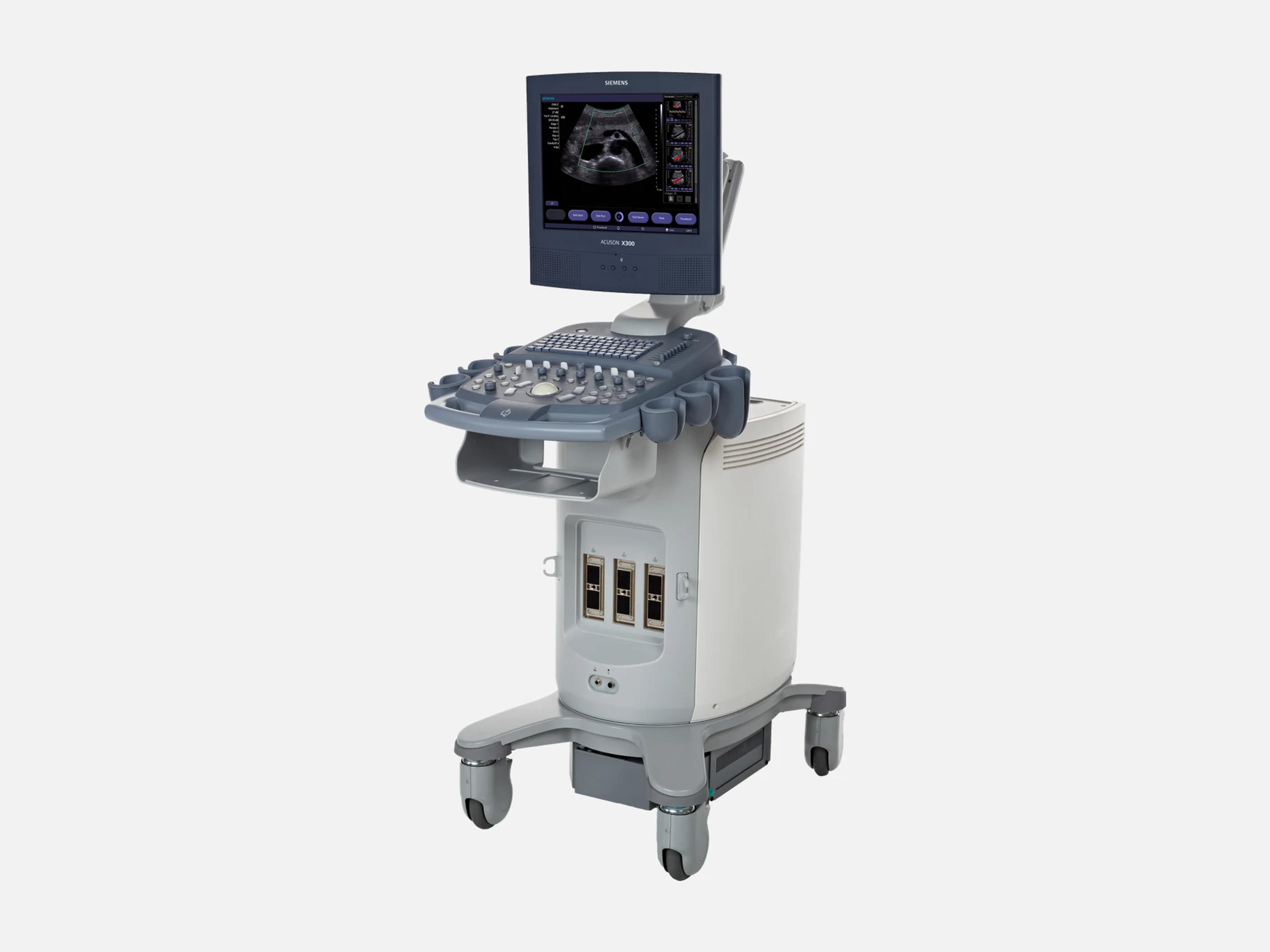
The Siemens ACUSON X300 is a cart-based color Doppler ultrasound system designed to support most multispecialty imaging. Its options add Stress Echo and Axius EF for cardiac work, Clarify VE and the syngo Arterial Health Package for vascular work, and OB/GYN reporting. It is distinct from the ACUSON X300 Premium Edition; Siemens aimed it at multi-use environments and the ACUSON X150 at private practices.
- Released
- 2006first FDA clearance
- Probe ports
- 2 active3 with Third Array Port
- Display
- 15-inchIPS flat panel, 1024 x 768
- Weight
- 102 kg225 lb
Compatible probes
Linear
- VF8-3 Needs the Third Array Port option
- VF10-5
- VF13-5
- VF13-5SP Intraoperative
Convex & microconvex
- CH5-2
- C8-5 Needs the Third Array Port option
Phased
- P4-2
- P8-4
Endocavity
- EV9-4 Endovaginal
- EC9-4
Specialty
- CW2 Doppler pencil
- CW5 Doppler pencil
- V5Ms Transesophageal (TEE)
Specifications
Display
- Display
15-inch high-resolution color flat panel, IPS, progressive scan, 1024 x 768 pixelsFlat Panel Display (FPD), 15-inch color, high resolution, progressive scan (non-interlaced), In Plane Switching (IPS) technology; resolution 1024 x 768 pixelsShow allShow less
- Monitor arm
Articulating; tilt 10 degrees up, 65 degrees down; swivel -80 to +80 degreesMulti-directional articulating monitor arm; monitor tilt 10 degrees up, 65 degrees down; swivel -80 to +80 degreesShow allShow less
Physical
- Weight
- 102 kg (225 lb); 98 kg without OEM devices
- Dimensions
- Height 137.9 cm (54.3 in); width 51.8 cm (20.4 in); depth 87.9 cm (34.6 in)
- Casters and wheel locks
Two front casters with bi-brake (direction, total lock); two rear with total lockWheel-lock mechanism; two front casters with bi-brake system (direction lock and total lock); two rear casters with total lockShow allShow less
- Control panel
HomeBase layout, back-lit, QWERTY keyboard; height adjusts 100 mm up/downControl panel with HomeBase layout; on/off task light and back-lit control panel; full size QWERTY keyboard; control panel height adjustment 100 mm up/down with lock lever; user-customizable control panelShow allShow less
- Transducer cables
- SuppleFlex cables with integrated cable management
- System design
- ErgoDynamic imaging system design
Power & battery
- Power
100-120/200-240 VAC, 50/60 Hz; built-in AC isolation transformerOne mainframe configuration for 100/115 V and 230 V environments; 100-120/200-240 VAC, 50/60 Hz; built-in AC isolation transformerShow allShow less
- Power consumption
- Maximum 600 VA with OEM devices; maximum heat output 2150 BTU/hr
Connectivity & options
- Transducer ports
Two active array ports plus a parking port; Third Array Port option adds a thirdTwo active array transducer ports plus one transducer parking port in the two-port configuration; the Third Array Port (option) adds a third array port, allowing up to three linear, curved and phased array transducers connected at once; electronic transducer selection. The VF8-3 and C8-5 need the third array port.Show allShow less
- Network and peripheral ports
Ethernet RJ45 (10/100 BaseT); two USB 2.0; two RS-232C serial; VGA out; foot switchOne Ethernet RJ45 (10/100 BaseT); two USB 2.0 ports; two RS-232C serial ports (D-SUB 9-pin); one VGA out (mini D-SUB 15-pin); one foot switch connectorShow allShow less
- DICOM
DICOM 3.0 option: storage, print, worklist, MPPS; OB/GYN, vascular, cardiac SR optionsDICOM 3.0 Connectivity (option): DICOM Storage Class User and DICOM Print Class User; DICOM Modality Worklist (option); DICOM MPPS, Modality Performed Procedure Step (option); DICOM OB/GYN, Vascular and Cardiac Structured Reporting (options)Show allShow less
- Export and network
TIFF, AVI and DICOM export; PACS/EMR connection over LAN or WLANExport formats TIFF, AVI and DICOM; PACS/EMR connection via LAN or WLAN; integration with DICOM-enabled networksShow allShow less
- Wireless data transfer (option)
- USB dongle to the facility LAN; IEEE 802.11n, 802.11g, 802.11b, 802.11a
- Removable media
Built-in DVD multi-drive (DVD-R/RW, CD-R/RW); USB flash drive exportBuilt-in DVD multi-drive (DVD-R/RW and CD-R/RW); USB flash drive export; exam archiving to CD/DVD and USBShow allShow less
- Hard drive and image storage
160 GB (100 GB for patient data); up to 150,000 images with compressionInternal 160 GB hard drive (100 GB reserved for patient data management); image storage up to 150,000 images with compression (B/W and color); DIMAQ-IP integrated workstation for patient information, images and reportsShow allShow less
- Documentation devices
Up to two on board: B/W printer and color printer/DVD recorder, in the cartUp to two on-board documentation devices (B/W printer and color printer/DVD recorder) integrated into the system cart and controlled from the control panelShow allShow less
- ECG / physiological interface
Standard 3-lead ECG; single and dual R-wave trigger; external ECG inputStandard 3-lead ECG interface with R-wave single and dual trigger, heart rate display and external ECG input selection; the Cardio-Vascular Package (option) also lists the physiological interface among its contentsShow allShow less
- Ultrasound System Security (option)
- Embedded virus protection
Imaging & software
- Imaging modes
2D, M, color M, color and power Doppler, PW and CW Doppler, DTI; duplex and triplex2D-mode in fundamental and harmonic (phase inversion and filtered); M-mode; Color M-mode; Color Doppler velocity mode; Power Doppler mode; Directional power Doppler; Pulsed Wave spectral Doppler (PW); Continuous Wave spectral Doppler (CW); DTI Doppler tissue imaging capability; Duplex and Triplex modes. The cleared device description also lists amplitude Doppler, combined modes, 3D, harmonic and 4D imaging.Show allShow less
- 2D frame rate
- Up to 498 fps, transducer dependent
- Beamformer
All-digital; up to 11360 processing channels; up to 4 transmit focal zonesAll-digital beam former; 2D-mode line density up to 256 lines; up to 11360 processing channels; up to 4 transmit focal zones; digital dynamic receive focusing; MultiBeam formation technologyShow allShow less
- Image depth (Release 2.5)
2–30 cm in 1.0 cm increments, transducer dependent; 30 cm with the CH5-2 and P4-22–30 cm in 1.0 cm increments, transducer dependent (maximum 30 cm with CH5-2 and P4-2; minimum 2 cm with VF13-5SP); 30 cm with the CH5-2 using SynApsShow allShow less
- MultiHertz multiple frequency imaging
- Up to five user-selectable transmit frequencies with the optional THI
- Tissue Harmonic Imaging (option)
- Phase inversion THI and filtering THI
- SynAps Synthetic Aperture Technology
- On the CH5-2, C8-5, VF8-3 and VF10-5; the user can turn it on and off
- Virtual Format Imaging
- Transducer dependent
- SieScape panoramic imaging (option)
B/W panoramic, images up to 60 cm long, up to 360 degrees of curvatureB/W panoramic imaging, images up to 60 cm long to a maximum curvature of 360 degrees; available on all curved and linear transducersShow allShow less
- SieClear compounding (option)
- Multi-view spatial compounding
- Clarify VE Technology (option)
- Uses energy Doppler flow information to enhance 2D-mode imaging
- Tissue Grayscale Optimization (option)
- One-button image optimization
- AutoColor Technology
- Automatic color Doppler optimization for different flow states
- 3-Scape real-time 3D (option)
Real-time 3D reconstruction during freehand acquisition; CH5-2 and EV9-4Real-time reconstruction of 3D images during freehand acquisition; available for the CH5-2 and EV9-4 transducersShow allShow less
- Doppler tissue imaging (option)
- Spectral and color DTI; requires the P4-2 or P8-4
- Cardiac imaging package
- Cardiac imaging modes, features and reporting tools
- Cardio-Vascular Package (option)
- Prerequisites for cardiac and certain vascular exams, with the physiological interface
- Stress Echo Imaging (option)
ECG-triggered loops; Protocol Editor up to 12 stages; 17-segment Wall Motion ScoringECG-triggered acquisition, display, comparison and archiving of cardiac loops; Protocol Editor up to 12 stages; Wall Motion Scoring, 17-segment model; integrated and user-programmableShow allShow less
- Axius EF (option)
- Calculates left ventricle volumes and ejection fractions
- syngo Arterial Health Package (option)
- Semi-automatic intima-media thickness (IMT) measurement
- Transducer technology
- Hanafy Lens
- QuickSet presets
- Up to 32 user-programmable system parameter presets
- User interface
- Windows-based; On-Screen Menu (OSM) for secondary imaging controls; Thumbnail Menu
- Revision and software
Versions 1.0 (February 2006), 2.0 (September 2007) and 2.5 (September 2014)Versions 1.0 (February 2006), 2.0 (September 2007) and 2.5 (September 2014), with DICOM conformance at V2.0 (September 2007); versions 3.0 (April 2008) through 7.0 (March 2011) are the ACUSON X300 premium edition'sShow allShow less
Environment
- Operating environment
+10°C to +40°C (50° to 104°F) without OEM devices; humidity 30–80% non-condensingAmbient temperature +10°C to +40°C (50° to 104°F) without OEM devices; humidity 30–80% non-condensing during operationShow allShow less
Compliance
- FDA 510(k)
K061946 (July 21, 2006); later K071036, K072676, K080760 and K093531K061946 (decision July 21, 2006; Class II); K071036 (May 16, 2007); K072676 (December 18, 2007); K080760, special 510(k) (April 25, 2008); K093531 (December 1, 2009)Show allShow less
- Design standards
- UL 60601-1, CSA C22.2 No. 601-1, EN/IEC 60601-1, 60601-1-1, 60601-1-2, 60601-2-37
- CE Mark
- Meets the Medical Device Directive
What to check when buying a used Siemens ACUSON X300
Reject probes with cracked housings, lenses or cables
A cracked or damaged housing, acoustic lens, strain relief or cable can compromise electrical safety and add cross-contamination risk, so such a probe must never be used.
How: Inspect each probe's housing, lens face, strain relief, cable and system connector under good light, and set aside any probe with a crack, split, cut or crushed section.
Check the software against the clip-carryover recall
On software 1.00.00 to 1.0.10 and 2.0.00 to 2.0.02, the system could show the previous patient's last captured clip in the next patient's study.
How: Read the installed software version, and on a unit in those ranges, ask for the record of the mandatory update under recall Z-2065-2011 and scan two test patients to confirm no clip carries over.
Confirm the mean pressure gradient fix was applied
On units with the cardiovascular options, the cardiac measurement package underestimated the mean pressure gradient traced from a stenotic Doppler waveform, which risks understating stenosis severity.
How: Ask for the service record of the correction under recall Z-1838-2011, which covered every X300 software version with those options installed.
Confirm the aortic stenosis calculation was corrected
On software revision 1.0.08, the system made incorrect calculations that could give inaccurate aortic stenosis estimates.
How: Read the software revision, confirm it is not 1.0.08, and ask for the record of the revised software installed under recall Z-0086-2009.
Common questions about the Siemens ACUSON X300
Is the Siemens ACUSON X300 FDA-cleared?
Yes. The ACUSON X300 Diagnostic Ultrasound System was cleared under 510(k) K061946, with a substantial-equivalence decision dated July 21, 2006, as a Class II device.
Later 510(k)s for the system are K071036 (May 16, 2007), K072676 (December 18, 2007), K080760 (April 25, 2008) and K093531 (December 1, 2009).
Is the ACUSON X300 the same as the ACUSON X300 Premium Edition?
No. The Premium Edition is a separate system with its own 510(k), K090276 (decided February 19, 2009), its own transducer list and its own software: versions 3.0 through 7.0, where the X300's are 1.0, 2.0 and 2.5.
The X300 has a 15-inch display, and the Premium Edition is specified with an 18-inch one. The Premium Edition's own transducer list includes the C6-2, C7F2, P5-1, P9-4, EV9F4 and BP9-4.
Does Siemens still support the ACUSON X300?
No. Siemens Healthineers has placed the ACUSON X300 on its US list of end-of-support ultrasound systems, and points owners to the ACUSON Juniper or ACUSON Maple as the upgrade.
What is the difference between the ACUSON X300 and the ACUSON X150?
They are separate systems: the X150 has its own 510(k), K081121, and Siemens aimed the X300 at multi-use environments and the X150 at private practices.
Several X300 probes also fit the X150: the CH5-2, P4-2, P8-4, VF10-5, VF13-5, EV9-4 and EC9-4.
Does the ACUSON X300 go by other names?
Yes. Its first 510(k), K061946, gives the device three proprietary names: Siemens Acuson X300 Diagnostic Ultrasound System, Sonoline Premier Plus Diagnostic Ultrasound System and Sonavista X 300 Diagnostic Ultrasound System.
The special 510(k) K080760 also names the SONOVISTA X300 ultrasound system among its devices.