Siemens ACUSON Juniper
Siemens Healthineers · ACUSON series
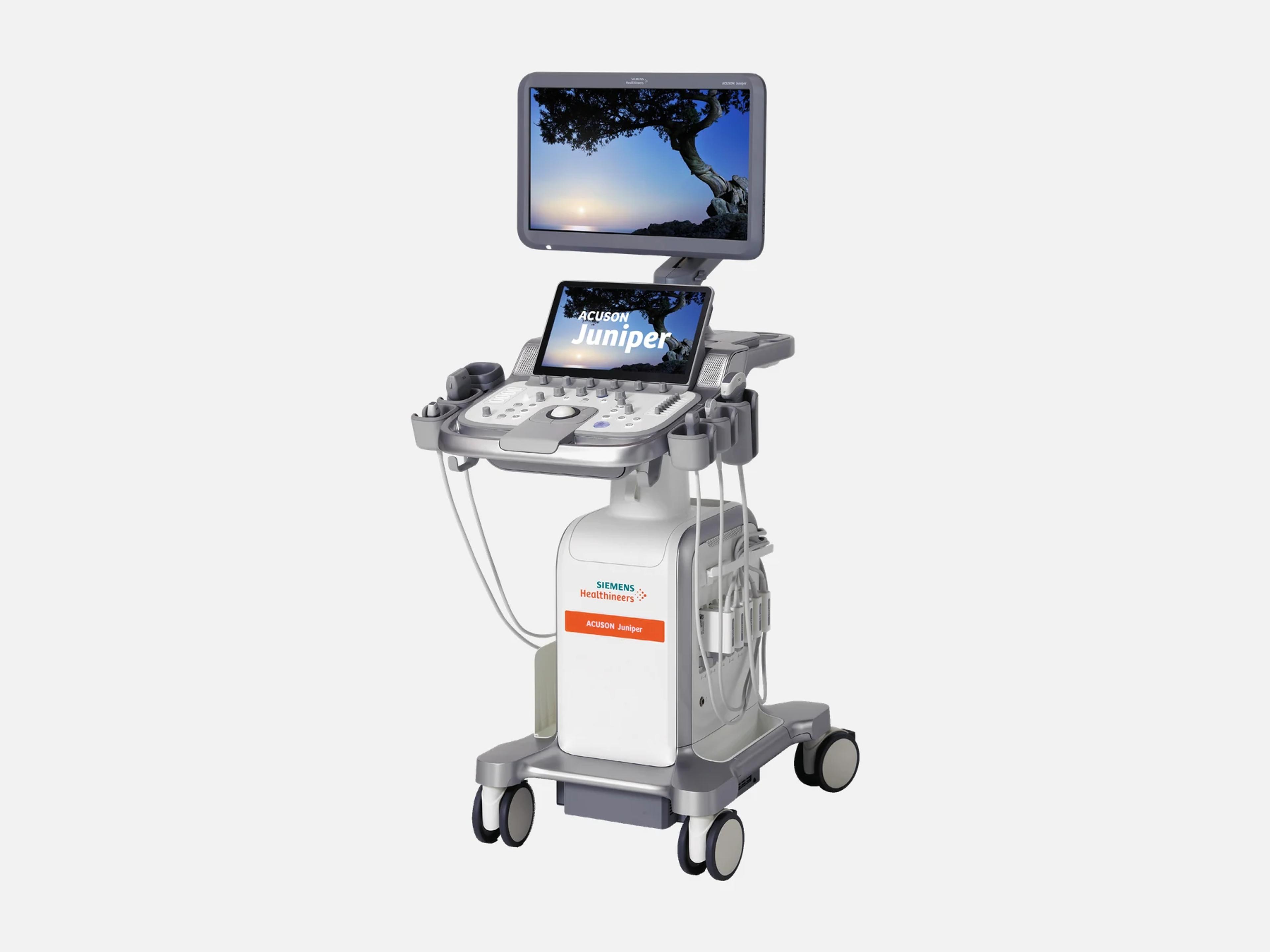
The Siemens ACUSON Juniper is a shared-service ultrasound cart for use across departments, designed to scan virtually every patient regardless of size, weight or condition. Its cardiac tools include eSie Measure and eSie Left Heart; Auto OB serves obstetric scanning, and UDFF and point shear wave elastography assess the liver. In the ACUSON range it sits below the Sequoia and Redwood and above the portable P500.
- Released
- 2018launched by Siemens
- Probe ports
- 5 activeplus 1 pencil CW port
- Display
- 23.6-inch21.5 in visible, full HD
- Weight
- 76 kg167.5 lb, without battery
Compatible probes
Linear
- 11L4 · 3.6–12.9 MHz
- 12L3 · 2.6–11.5 MHz
- 13L4 · 3.9–12.6 MHz
- 14L4 · 4–12.7 MHz Introduced with Juniper 2.0
- 16L4 · 4.3–15.4 MHz
- 18H5 · 4.5–18 MHz Hockey stick
Convex & microconvex
- 5C1 · 1.4–5 MHz
- 6C1 · 1–5.6 MHz Single crystal; introduced with Juniper 2.0
- 7C2 · 2.1–7 MHz
- 11M3 · 3.5–11 MHz Micro-convex
- 9VC2 · 1.8–8.5 MHz Curved volume (4D)
- 8VC3 Curved volume (legacy)
Phased
- 5P1 · 1.1–5 MHz Single crystal
- 8V4 · 2.7–8 MHz Vector
- 10V4 · 3.4–10.4 MHz Vector
Endocavity
- 9MC3 · 3.2–8.5 MHz Micro-curved
- 10MC3 · 3.5–10.2 MHz Micro-curved
- 9VE4 · 3.1–8.7 MHz Endocavity volume (4D)
- 9VE3 · 3–9 MHz Endocavity volume (legacy)
Specialty
- 5VT · 3.1–9.2 MHz Transesophageal
- 10BCC3 · 3–10.4 MHz Urology
- CW2 2.0 MHz CW pencil
- CW5 5.0 MHz CW pencil
- CW8 8.0 MHz CW pencil; not sold in the U.S. at Release 2.5
Specifications
Display
- Monitor
23.6 in (59.9 cm) full HD LCD, LED backlit; 21.5 in visible; IPS, 1920 × 1080, 1000:1Physical size 23.6 in (59.9 cm), visible size 21.5 in; full HD 1080p (1920 × 1080) flat-panel LCD with LED back-lighting and IPS technology; contrast ratio 1000:1.Show allShow less
- Touch screen
- 13.3 in LCD touch panel, 16:9, 1920 × 1080, tiltable, with two USB ports
- Monitor articulation
- ±180º left/right swivel; upright height 163–141 cm; folds down for transport
Physical
- Weight
- 76 kg (167.5 lb) without Battery Support; 78.2 kg with it
- Dimensions
Width 50.8 cm, height 122.5–163.4 cm, depth 65.4 cm (84.5 cm max)Width 50.8 cm (1.6 ft); height 122.5 to 163.4 cm (4.02 to 5.4 ft); depth 65.4 cm (2.14 ft). Maximum depth 84.5 cm; minimum fold-down system height 122 cm.Show allShow less
- Transducer ports
- 5 active, plus 1 pencil (CW) port; Twin Cam zero-insertion-force connectors
- Controls
Floating panel, ±90º swivel, 11 cm height travel; backlit keys; laser-optical trackballFloating control panel with left/right swivel of ±90º and 11 cm vertical articulation; Off/White/Blue control backlighting; laser-optical trackball; touch-screen keyboard with an optional pull-out keyboard; customizable height, angle and keys.Show allShow less
- Cart
Front and rear handles; cable hooks; 7 transducer cup holders plus specialty holdersFront and rear handles; multiple cable management hooks; 7 transducer cup holders and specialty holders for endocavity, intra-operative, Aux CW and 4D transducers; rear storage basket and magnetic side storage bin.Show allShow less
- Gel warmer
- Integrated, standard: 31, 34 and 37 °C; larger 1-liter gel holder optional
- Fan noise
- 28 to 33 dBA
Power & battery
- Battery Support
- Option: replaceable, rechargeable lithium battery with Battery Health Management
- Battery time
- Scanning up to 75 min; operation up to 90 min (standard environmental conditions)
- Power
100–240 Vac, 50/60 Hz (90–264 Vac universal input); max 1.0 kVA; max heat 2400 BTU/hr100 to 240 Vac (50/60 Hz), universal input 90 to 264 Vac; maximum power consumption 1.0 kVA, which may vary with configuration; maximum heat output 2400 BTU/hr.Show allShow less
- Power consumption at 115 V (2019)
- Scanning 0.281 kW, freeze 0.242 kW, hibernate/standby 0.119 kW, off 0.039 kW
- Boot time from off
About 68 seconds, depending on security settings (Release 2.5)Approximately 68 seconds from off, depending on security settings (Release 2.5). In 2019: power-on 2 min 4 s with virus protection on and 1 min 36 s with it off; power-off 47 s.Show allShow less
- QuikStart Standby Mode
- Option: sleep in 15 s, on from sleep in about 15 s; transport mode up to 180 minutes
Connectivity & options
- DICOM
DICOM 3.0: storage, print, commitment, worklist, MPPS, query/retrieve, SRDICOM 3.0: Storage and Print Class User, storage commitment, Modality Worklist, MPPS, and Structured Reporting for Vascular, OB/GYN and Cardiac (Release 2.5). Verification, Storage, Storage Commitment, Query, Retrieve, Worklist, MPPS and Print application entities (VB10 and VC10), plus Comprehensive SR (VC10).Show allShow less
- IP support
- IPv4 only; IPv6 not supported (VC10)
- Wireless Data Transfer
Option: internal Wi-Fi, IEEE 802.11a/b/g/n/ac, WPA/WPA2 personal and enterpriseOption: internal Wi-Fi module, IEEE 802.11a/b/g/n/ac, WPA/WPA2 personal and enterprise; supports DICOM worklist, storage commitment and store, and Smart Remote Services.Show allShow less
- Input/output ports
USB 2.0 ×2 and USB 3.0 ×2 (USB-A), Gigabit Ethernet (RJ45), S-Video, DVI, HDMIUSB 2.0 ×2 and USB 3.0 ×2 (USB-A), Gigabit Ethernet (RJ45), S-Video, DVI and HDMI; two USB 2.0 ports beside the touch screen and four on the back.Show allShow less
- Data export
- PDF, TIFF, AVI, JPG and DICOM; to PACS or EMR over LAN or WLAN
- Documentation devices
Up to 2 on-board devices: B/W and color printers, DVD recorderUp to 2 on-board documentation devices integrated into the system cart; supported devices include the UP-D898 and UP-D711 B/W printers, the UP-D25MD color printer and the UR-50BD-SK DVD recorder.Show allShow less
- ECG and physio
Standard 3-lead ECG; auxiliary physio input/output; Physio Module option; 2 physio portsStandard 3-lead ECG interface with auxiliary input/output for third-party physiology signals; Physio Module (option); 2 physio ports (Release 3.0).Show allShow less
- Barcode Reader
- Option, USB-attached; reads 1D and 2D patient barcodes
- Dual USB Foot Switch
- Option, programmable
- Protective Control Panel Cover
- Option: reusable cover for control panel and touch screen, for infection control
- Remote service
Kinectus Remote Service; Smart Remote Services (SRS); eSieLink Remote AssistanceKinectus Remote Service, a cloud-based connection platform delivering software updates; Smart Remote Services (SRS); eSieLink Remote Assistance (Remote Assist, Remote Trainer) for remote application support.Show allShow less
- Security
Cybersecurity management system; virus protection; Data Storage Encryption (option)Cybersecurity management system; embedded virus protection; Data Storage Encryption (option), protecting patient information and system settings stored on the system, with recovery from a USB storage device holding the recovery key.Show allShow less
Imaging & software
- Revision and software
- Release 3.0 (VC10); earlier 1.0 (VA10), 1.5 (VB10), 2.0 (VB11), 2.5 (VB30)
- Imaging modes
2D and THI; color, power and directional power Doppler; PW, steerable CW, aux CW; M-mode2D: Fundamental 2D, Phase Inversion THI, Filtered THI, Alternative THI. Color Doppler: velocity-based color Doppler, Power Doppler, Directional power Doppler, Color Doppler tissue imaging. Spectral Doppler: Pulsed wave, Steerable Continuous Wave, auxiliary continuous wave on the pencil probe, Spectral Doppler tissue imaging, Duplex and Triplex. M-mode: M-mode, Color M-mode, Anatomical M-mode. Cleared modes also include High PRF Pulsed Wave Doppler, 3D/4D volume imaging, 2D with elastography and 2D with contrast agent imaging.Show allShow less
- Frequency range
- 1–20 MHz
- Processing channels
- Up to 3,670,016
- Processor and memory
- V1000 processor, 4 × 3.35 GHz; 16 GB DDR4 RAM; 3150 MB cine memory
- Operating system
- Windows 10
- Storage
- 500 GB internal SSD; Blu-ray/DVD/CD drive (reads Blu-ray, writes DVD±R and CD-R)
- Image optimization
eSieImage Multiparametric Optimization; Auto TEQ brightness optimization; UltraArteSieImage Multiparametric Optimization, adjusting imaging parameters automatically while scanning; Auto TEQ, optimizing image brightness across the field of view; UltraArt universal image processing.Show allShow less
- Spatial compounding
SieClear Multi-view; Advanced SieClear, also in Color and Power Doppler, up to 7 anglesSieClear Multi-view Spatial Compounding; Advanced SieClear Spatial Compounding, including in Color and Power Doppler, with up to 7 steering angles on linear and on curved array transducers.Show allShow less
- Image processing
Dynamic TCE speckle reduction; Dynamic Persistence; Custom Tissue Imaging; MultiHertzDynamic TCE post-processing speckle reduction; Dynamic Persistence for B-mode and Color, against ghosting when probe or patient motion is detected; Custom Tissue Imaging, adjusting the speed of sound; MultiHertz, up to seven 2D and THI frequencies and up to three color and spectral Doppler frequencies per transducer.Show allShow less
- Color artifact suppression
Auto Flash Color Artifact Suppression, with no user interactionAuto Flash Color Artifact Suppression detects motion artifacts from probe and patient movement and reduces color artifacts dynamically, with no user interaction.Show allShow less
- Vascular
Clarify VE (7 levels); syngo Arterial Health Package for carotid IMT (option)Clarify Vascular Enhancement (VE), real-time contrast resolution and border detection of vascular structures, with 7 user-selectable levels; syngo Arterial Health Package (AHP) (option), Carotid Intima-Media Thickness measurement with optional reference to published normative tables.Show allShow less
- Cardiac
eSie Measure, eSie Left Heart, syngo VVI, Stress Echo (options); DTI; LVO contrasteSie Measure (option), 1-click cardiac measurements for 2D, M-mode and Doppler; eSie Left Heart (option), EF, EDV and ESV for LV and LA; syngo Velocity Vector Imaging (option), GLS, GCS and GRS; Stress Echo (option), with 16/17 segment wall motion scoring; DTI Velocity and DTI Energy; Left Ventricular Opacification for endocardial definition.Show allShow less
- OB/GYN
Auto OB (BPD, OFD, HC, AC, HL, FL) and Auto Follicle (options); LightSource RenderingAuto OB (option), automated biometric measurements of BPD, OFD, HC, AC, HL and FL; Auto Follicle (option), automated follicle measurements; LightSource Rendering for 3D/4D fetal visualization with the 9VC2.Show allShow less
- 3D and 4D
4D Imaging (option), fourSight, up to 19.3 vol/s on 9VC2, 9VE4; 3-Scape freehand 3D4D Imaging (option) with fourSight 4D transducer technology, up to 19.3 vol/sec, on the 9VC2 and 9VE4; Advanced fourSight Technology with MultiSlice up to 36 slices; Thick Slice Imaging and Curved MPR; 3-Scape freehand 3D acquisition on the 5C1, 6C1, 7C2, 9MC3, 10MC3, 9VC2 and 9VE4.Show allShow less
- Elastography
eSieTouch Elastography; eSie Touch Elasticity Imaging; pSWE; Virtual Touch QuantificationeSieTouch Elastography, qualitative elasticity imaging for breast lesion characterization; eSie Touch Elasticity Imaging (option), relative tissue stiffness with area, distance and strain ratio measurements; Point Shear Wave Elastography (pSWE) for liver stiffness; Virtual Touch Quantification (option), ARFI-based, with the 5C1 and 6C1.Show allShow less
- Liver fat
- Ultrasound-derived fat fraction (UDFF) to quantify liver steatosis
- Contrast
Contrast Enhanced Ultrasound (option) on the 6C1 and 14L4; Burst and Flash SequencingContrast Enhanced Ultrasound (option) with Burst and Flash Sequencing destruction techniques, on the 6C1 (abdomen, abdomen-difficult, renal; focal liver lesions) and the 14L4 (breast, thyroid).Show allShow less
- Needle guidance
- Enhanced Needle Visualization (option), all linear transducers, in-plane angles up to 35°
- Panoramic
- SieScape (option), extended field of view up to 240 cm long or 180º
- Measurement and workflow
eSie Calcs tracing; eSieScan protocols (option); Exam Restart; full screen modesyngo eSie Calcs native tracing (area, maximum diameter and volume); eSieScan Workflow Protocols (option) for Cardiac, Vascular, OB and Breast exams; Exam Restart adds images to an already closed exam, with a 24-hour limit; full screen mode enlarges the clinical image up to 56%.Show allShow less
Environment
- Operating
- +10 to +40 °C (50–104 °F); 10–80% RH non-condensing; 700–1060 hPa (3000 m)
- Storage and transport
- -20 to +60 °C; 10–95% RH non-condensing
Compliance
- FDA 510(k)
K230207 (February 2023), K221190, K201130K230207 (February 2023): added the 9VC2, 9VE4, L10-5v and P4-2 transducers and the Lightsource application, renamed from Amnioscopic rendering. K221190: added the 6C1, 14L4 and CW8 transducers, a battery pack, a B/W printer and Contrast Agent Imaging. K201130: added the 13L4 and 5VT transducers.Show allShow less
What to check when buying a used Siemens ACUSON Juniper
Inspect the transducer ports for fluid and residue
The transducer ports must never be cleaned with gauze, cloths or solutions, and spray cleaners or poured fluid must never reach the system.
How: Look into each port and over the surrounding surfaces for fluid ingress or cleaning residue, and ask whether the gel warmer was ever immersed.
Confirm the clip-storage recall fix on Juniper 1.0
On Juniper 1.0 software VA10D, VA10E and VA10F, the clip store function fails when the system has a disk-full error, the fault behind FDA recall Z-0748-2022.
How: Read the installed software version, confirm that a VA10D, VA10E or VA10F unit received the VA10G correction, and check the free storage.
Confirm Battery Support and read the battery status
Battery Support, a replaceable and rechargeable lithium battery with Battery Health Management, is an option and is not fitted to every unit.
How: Confirm the unit has Battery Support, then read the on-screen battery information.
Confirm which options are licensed on the unit
Features such as 4D Imaging, Virtual Touch Quantification, eSie Left Heart, eSie Measure, Stress Echo, Contrast Enhanced Ultrasound and Wireless Data Transfer are sold as options.
How: List the options licensed on the system, and demonstrate each option in the seller's quote with its own controls.
Common questions about the Siemens ACUSON Juniper
Is the Siemens ACUSON Juniper FDA-cleared?
Yes. It is FDA-cleared under 510(k). K201130 added the 13L4 and 5VT transducers; K221190 added the 6C1, 14L4 and CW8 transducers, a battery pack and contrast agent imaging; and K230207, from February 2023, added the 9VC2, 9VE4, L10-5v and P4-2 transducers.
The CW8 was not for sale in the U.S. at Release 2.5.
Does the Siemens ACUSON Juniper do contrast-enhanced ultrasound?
Yes, as an option. Contrast Enhanced Ultrasound runs on the 6C1 for abdominal and renal studies and on the 14L4 for breast and thyroid studies, and Left Ventricular Opacification supports endocardial definition.
Contrast agent imaging was added under 510(k) K221190. At Release 2.5, the FDA had cleared ultrasound contrast agents only for left ventricular opacification, the liver and vesicoureteral reflux.
What is the ACUSON Juniper CV Edition?
ACUSON Juniper, CV Edition is a configuration of the ACUSON Juniper that combines its available cardiovascular options into a cardiology system.
Is the ACUSON Juniper an upgrade from the ACUSON X300 or Antares?
Yes. The ACUSON Juniper is an upgrade path for both the ACUSON Antares and the ACUSON X300 at the end of their support.
Where is the Siemens ACUSON Juniper made?
Its manufacturer of record is in Issaquah, Washington, and its manufacturing sites include Seongnam-si in the Republic of Korea and Kosice in Slovakia.