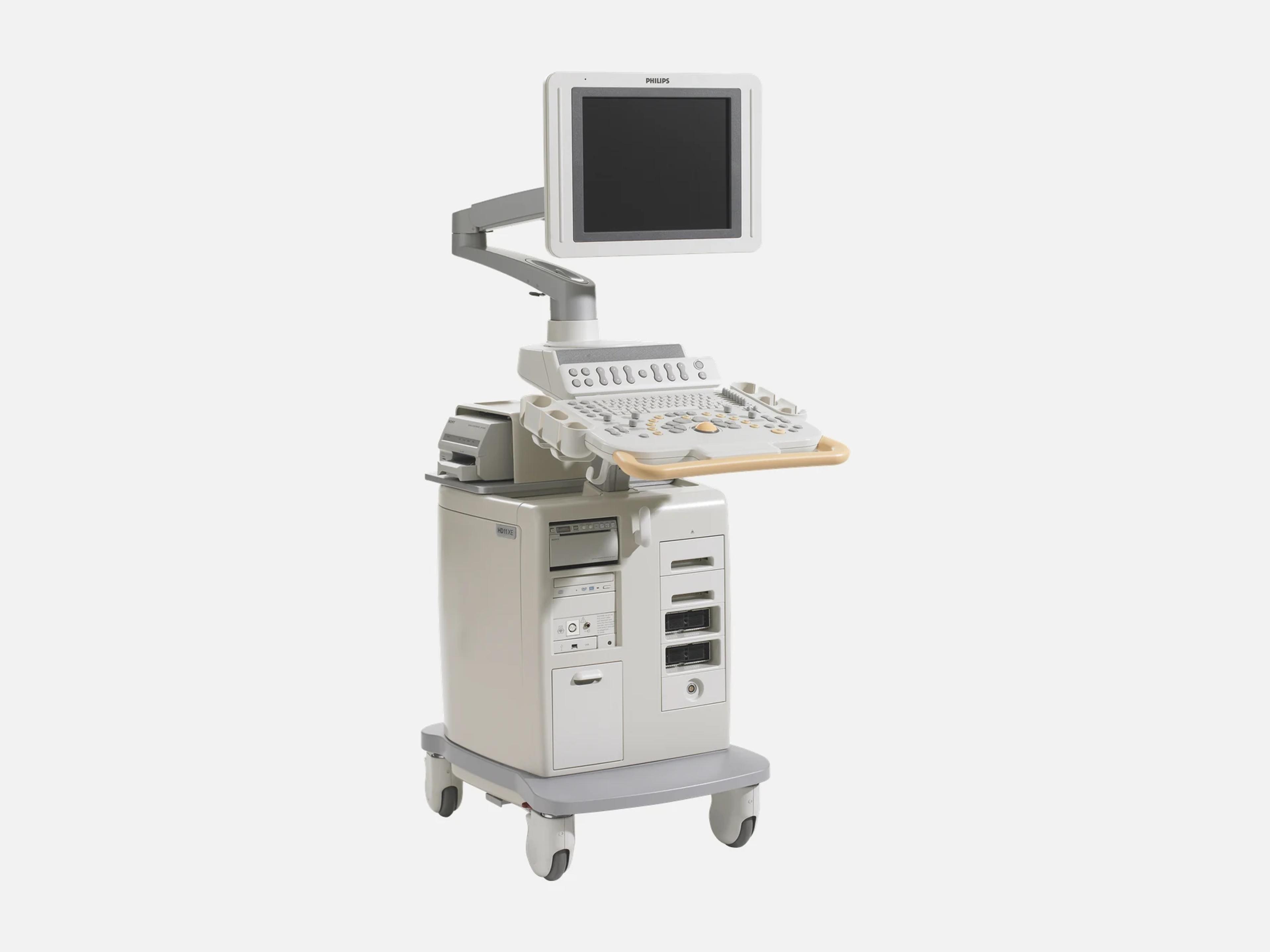
The Philips HD11 XE is a cart-based ultrasound system for public and private hospitals, satellite clinics and specialized practices. Standard modes include 2D with Pulse Inversion Harmonic Imaging, Adaptive Color Doppler and Tissue Doppler Imaging; options add stress echo, TEE, 4D Imaging and 3D Fetal Echo STIC. It is the newer version of the HD11, with a flat-panel monitor and expanded cardiac capabilities.
- Released
- 2005launched by Philips
- Probe ports
- Up to fiveCartridge, Explora, Pencil
- Display
- 51 cmTFT LCD monitor
- Weight
- 100 kg220 lb, without peripherals
Compatible probes
Linear
- L12-5 50 mm
Convex & microconvex
- C6-3 Intercostal access; renamed from C5-2
Phased
- S3-1 Sector; contrast LVO, and transcranial with the D2tcd
Specialty
- V6-2 Volume transducer; renamed from 3D6-2
- V8-4 Volume transducer; renamed from 3D8-4
- L15-7io Intraoperative; vascular imaging
- BP10-5ec Biplane
- D2tcd · 2–2 MHz Doppler pencil; transcranial with the S3-1
- S7-2omni TEE with harmonic imaging
- S7-3t TEE for pediatric and small adult patients
- OmniPlane III TEE, model 21378A, through the OmniPlane Adapter
- ViewFlex catheter Third-party ICE catheter; select geographies
- ViewFlex Plus ICE Catheter Through the Patient Isolation Module
Specifications
Display
- Display
- 51 cm TFT LCD flat panel
- Resolution
- 1280 × 1040 (monitor)
Physical
- Weight
- 100 kg (220 lb) without peripherals
- Dimensions
- Width 53.3 cm; depth 110.5 cm; height 151.1–173.9 cm, adjustable
- Probe ports
Up to five transducer connections: Cartridge, Explora and Pencil connector stylesConfigurable for up to five transducer connections, in Cartridge, Explora and Pencil connector styles; the legacy T6H transducer needs an adapter.Show allShow less
- Controls
Height-adjustable monitor and control panel, integrated footrest, QWERTY keyboardMonitor and control panel independently height-adjustable; integrated footrest; QWERTY keyboard.Show allShow less
Power & battery
- Power
- 100–240 V AC, 50/60 Hz; 750–1000 VA, depending on system configuration
- Power protection
- Uninterrupted power supply (UPS)
Connectivity & options
- DICOM
DICOM export to removable media; DICOM Networking option for print, store and worklistThe base system exports DICOM studies to removable media. The DICOM Networking option adds DICOM Print and Store, Modality Worklist, Performed Procedure Step and Structured Reporting, and works with most DICOM management systems; storage commitment and verification are supported (Release 1.1.x.x). Structured Reports export only for obstetric, gynecology and cardiac studies. Remote service network.Show allShow less
- Removable media
Release 1.1.x.x: CD-R, CD-RW, MOD; Release 2.0 adds DVD-R, DVD-RW and USBRelease 1.1.x.x: CD-R, CD-RW or MOD. Release 2.0: CD-R, CD-RW, DVD-R, DVD-RW, USB or MOD. Multi-session CD and optional peripheral devices.Show allShow less
- Internal storage
- Minimum 120 GB hard-drive space
- Options
QLAB, stress echo, contrast, TEE, 4D Imaging, 3D Fetal Echo STIC, iSlice, Panoramic…QLAB advanced quantification; stress echo; contrast imaging (Left Ventricular Opacification); TEE; 4D Imaging; 3D Fetal Echo STIC; iSlice; Panoramic Imaging, for an extended field of view; QLAB IMT plug-in; DICOM Networking; intracardiac echo (ICE) with the third-party ViewFlex catheter, in select geographies only; clinical packages for anesthesia and emergency.Show allShow less
Imaging & software
- Imaging modes (standard)
2D, Adaptive Color Doppler, Color Power Angio, PW and CW Doppler, TDI, Anatomical M-modeStandard on every system: 2D with Pulse Inversion Harmonic Imaging; Adaptive Color Doppler with Color Power Angio; pulsed wave and continuous wave Doppler with Adaptive Doppler technology and high PRF; Tissue Doppler Imaging (TDI), including Color TDI and pulsed wave TDI; Anatomical M-mode.Show allShow less
- Image processing
SonoCT compound imaging, XRES adaptive image processing, Fusion key, trapezoidal formatSonoCT compound imaging (transmit beam-steering); XRES adaptive image processing; Fusion key with Resolution, General and Penetration settings, integrating low- and high-frequency ranges in one image; trapezoidal image format.Show allShow less
- Beamformer
9,216 digitally processed channels; broadband beamforming9,216 digitally processed channels; broadband transducers and beamformer capturing the full bandwidth of ultrasound signals.Show allShow less
- Automation
iSCAN, Intelligent Doppler, High Q Automated Doppler Analysis, customizable presetsiSCAN: one-button gain, TGC and compression on 2D vascular images, and automatic scale and baseline in Doppler modes; Intelligent Doppler for vascular scanning-angle adjustment; High Q Automated Doppler Analysis for measurements from user-defined Doppler waveforms; presets in every clinical software package, customizable.Show allShow less
- QLAB (option)
On-cart and off-cart; plug-ins 2DQ with Color Kinesis, Strain Quantification, IMT, ROIQLAB on-cart and off-cart (on Xcelera). Plug-ins: Cardiac 2D quantification (2DQ) of chambers and vessels, with Color Kinesis and Mitral Annular Color Kinesis; Strain Quantification (SQ) of tissue velocity, strain rate and strain; Automated Intima Media Thickness (IMT) of carotid and other superficial arterial walls; Region of Interest (ROI) quantification of pixel intensities. QLAB supports the iSlice display, with automated stacked contours.Show allShow less
- Stress echo (option)
Fixed and user-defined protocols, up to eight stages and eight views; wall motion scoringIntegrated into the user interface; fixed and user-defined protocols capturing up to eight stages and eight views; single-, quad- and multi-cycle acquisition; left ventricular opacification (contrast) with stress; wall motion scoring and reporting.Show allShow less
- Volume imaging
4D Imaging, iSlice, thick slice, 3D Fetal Echo STIC, freehand 3D with multiplanar views4D imaging with volume acquisition and display, and simultaneous visualization and measurement in three planes; iSlice and thick slice; 3D Fetal Echo STIC; qualitative freehand 3D imaging with multiplanar views.Show allShow less
- Study review and reports
Image thumbnails for study building and exam status; exam reports with embedded imagesOn-screen image thumbnails for study building and exam status; exam reports with embedded images; one-touch on-line Help.Show allShow less
- Revision and software
R1.1.x.x, R1.2.x and R2.0, the latest HD11 XE releaseRelease 1.1.x.x, Release 1.2 (R1.2.x) and Release 2.0, the latest HD11 XE release; their DICOM conformance statements are dated 2005-11-08, 2006-10-06 and 2007-08-01. The HD11's own releases, R1.0.1 to R1.0.8, are a separate line.Show allShow less
Compliance
- FDA 510(k)
K062247 (2006-08-18); K043535 (2005-01-06), as the HD11 Diagnostic Ultrasound SystemK062247, Special 510(k), decided 2006-08-18, for the C6-3, V8-4 and V6-2 transducers and the ViewFlex catheter; K043535, Traditional 510(k), decided 2005-01-06. Both records name the device HD11 Diagnostic Ultrasound System.Show allShow less
What to check when buying a used Philips HD11 XE
Check recall Z-0987-2007 and its software correction
On units with serial numbers on the recall list, exiting an imaging mode while Active Annotation was in use could leave the system in an undefined state where the image scale may not match on-screen measurements and graphics.
How: Compare the unit's serial number with the recall list and confirm the corrective software release is installed.
Watch a cold power-on for smoke
An FDA malfunction report describes smoke coming out of an HD11 XE when it was turned on.
How: Ask to see the system started from cold, and stop the purchase if any smoke appears.
Check any S7-3t TEE probe against recall Z-1842-2020
Some S7-3t and S8-3t probes were programmed incorrectly in manufacturing, so the system cannot measure distal-tip temperature correctly and auto-cool may not start, which could injure a patient.
How: Ask whether the probe was remediated, because affected probes cannot be detected on HD11 systems and the recall tells HD11 users to stop using them; its records name the HD11, not the HD11 XE.
Have each TEE probe safety-tested on the system
A stand-alone tester only indicates leakage on a TEE probe, and any failure on it calls for complete safety testing of the probe with the ultrasound system.
How: Ask a biomed to leakage-test every TEE probe offered with the unit and, if one fails, to run the complete safety test with the system.
Common questions about the Philips HD11 XE
Is the Philips HD11 XE FDA-cleared?
Yes. The HD11 XE is FDA-cleared under 510(k) K062247, a Special 510(k) decided on August 18, 2006, which includes the C6-3, V8-4 and V6-2 transducers and the ViewFlex catheter. The 510(k) records name the device HD11 Diagnostic Ultrasound System, which was also cleared under K043535, a Traditional 510(k) decided on January 6, 2005.
Do iE33 and EnVisor transducers work on the HD11 XE?
Some do. Select transducers are compatible across Philips platforms, including the iE33 and EnVisor, and the HD11 XE supports many from the Explora line. It takes sector, linear-array, curved linear array, volume, TEE and Doppler pencil transducers with frequencies from 1 to 15 MHz, in Cartridge, Explora and Pencil connector styles. The legacy T6H needs an adapter, and the OmniPlane III TEE probe connects through the OmniPlane Adapter.