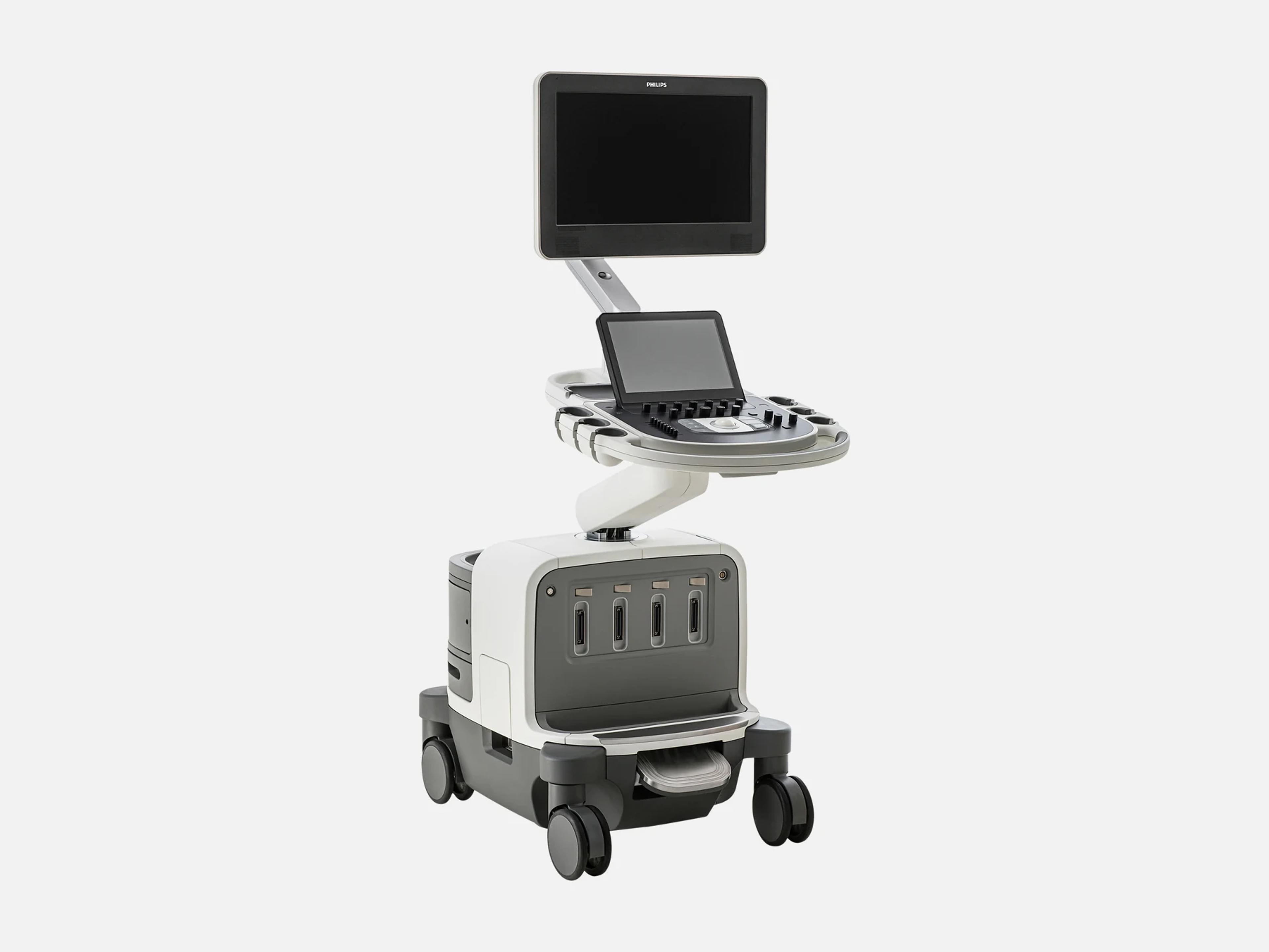
The Philips EPIQ 7 is a premium cart-based ultrasound system for obstetrical, gynecological, cardiology, vascular and general imaging practices. Its designations include the EPIQ 7C, optimized for cardiology, and the EPIQ 7G, for general imaging, shared service and radiology. On the EPIQ 7, one xMATRIX transducer runs 2D, 3D/4D, Live xPlane and Doppler; on the EPIQ 5, xMATRIX is offered as an upgrade path.
- Released
- 2013launched by Philips
- Probe ports
- 4on Release 1.0 systems
- Display
- 21.5-inch54.6 cm LCD, articulating
- Weight
- 104.3 kg230 lb
Compatible probes
Linear
- L12-3
- L12-3ERGO
- L12-5 50 50 mm; breast and small-parts fusion
- L15-7io Intraoperative
- L18-5
- eL18-4 Standard and electromagnetic variants
- VL13-5 Volume linear; used with VPQ for carotid plaque
Convex & microconvex
- C5-1 PureWave curved array; large abdomens
- C9-2 PureWave curved array; high-frequency OB
- C8-5
- mC7-2 Added in the EPIQ 4.0 submission, K182857
- X6-1 PureWave xMATRIX; abdominal and OB volume imaging
- V6-2 Motorized 3D volume
Phased
- S5-1 PureWave sector; difficult cardiology and transcranial
- S8-3
- S12-4
- X5-1 xMATRIX; adult cardiology
- X7-2 PureWave xMATRIX
Endocavity
- C10-3v PureWave endocavity; fibroid and complex ovarian cases
- C10-4ec Prostate fusion
- 3D9-3v
Specialty
- S7-3t Transesophageal
- X7-2t xMATRIX transesophageal
- D2cwc Non-imaging CW pencil probe
- D5cwc Non-imaging CW pencil probe
- D2tcd Non-imaging pencil probe
- VeriSight Intracardiac echo catheter; EPIQ 7C only
- VeriSight Pro Intracardiac echo catheter; EPIQ 7C only
Specifications
Display
- Display
- 21.5-inch (54.6 cm) LCD monitor; 720° of articulation; folds down for transport
- Touch screen
- 12-inch touch control panel with soft-key controls, beside physical knobs and buttons
- MaxVue
- High-definition display mode for dual, side/side, biplane and scrolling imaging modes
Physical
- Weight
- 104.3 kg (230 lb)
- Dimensions
- Width 60.6 cm; height 146–171.5 cm; depth 109.2 cm
- Transducer ports
- Four imaging-transducer receptacles on Release 1.0 units; one active at a time
- Control panel
Articulating, with 180° of movement and 25.4 cm of height adjustmentArticulating control panel with 180 degrees of movement and 25.4 cm of height adjustment; ambient lighting gives visual cues for the keyboard and transducer ports; a button on the system handle locks and unlocks the control panel arm.Show allShow less
- Transport
Brake, system and back handles, fold-down monitor, cable hooks, catch tray, sleep modeCart with a system brake, a system handle and back handles on the control panel for pushing; the monitor folds down to reduce height for transport; integrated cable hooks and catch tray; sleep mode lets the system be moved and booted up again in seconds.Show allShow less
- Operating noise
- 37–41 dB in manufacturer noise testing
Power & battery
- Power
- 100–240 Vac, 50 or 60 Hz, 600 VA
- Battery
- Release 1.0: battery-powered Sleep Mode; no scanning on battery
Connectivity & options
- DICOM services
Worklist, image and SR storage, storage commitment, MPPS, query/retrieve, networked printDICOM worklist; storage of images and structured reports to the network; storage commitment; media storage to CD, DVD or USB; networked DICOM print; query/retrieve; MPPS. On Release 1.8: Comprehensive SR Storage, Modality Worklist, Modality Performed Procedure Step and Storage Commitment.Show allShow less
- Netlink DICOM 3.0
- Purchasable option required for DICOM network services; DICOM printing needs no option
- Query/retrieve
Review of CT, NM, MR, mammography and ultrasound images; requires a Query Retrieve optionQuery/retrieve of DICOM images (CT, NM, MR, mammography, ultrasound) for review on the system, with the Ultrasound Query Retrieve or Multi-Modality Query Retrieve option.Show allShow less
- Network
Wired 10/100/1000 BaseT (RJ-45); wireless 802.11 g/n/ac, optional or internal adapterWired Ethernet 10/100/1000 BaseT (RJ-45); wireless 802.11 g/n/ac through the optional adapter or an internal wireless adapter (Release 4.0.x and 7.0.x); the wired network has priority when both are configured.Show allShow less
- Wireless DICOM
- Supported; availability varies by geography
- Storage and media
Local repository; network, DVR, CD, DVD and USB; JPEG, AVI and DICOMLocal clinical data repository, with patient data on a separate partition; off-line storage to the network, DVR, DVD and USB; removable-media exchange on CD, DVD or USB (Release 4.0.x); images stored as JPEG, AVI and DICOM.Show allShow less
- MPR DICOM Export
- Sends 3D MPRs in the X, Y and Z planes to PACS
- Remote services
iSSL connection, proactive monitoring, remote desktop and online support requestsiSSL secure connection to the Philips remote services network over the site's internet access; proactive remote detection of anomalies by Philips service; remote desktop (Virtual Visit) for technical and clinical troubleshooting, availability varying by geography; online support requests entered from the system.Show allShow less
- Security
- Government Security, Security Plus, SafeGuard
Imaging & software
- Imaging modes
2D, 3D/4D, Live xPlane, Live MPR, MPR, Doppler, color Doppler, CPA; elastography, CEUS2D, 3D/4D, Live xPlane (two full-resolution planes acquired simultaneously), Live MPR, MPR, Doppler, color Doppler and CPA, all on a single xMATRIX transducer; plus MicroCPA, strain and shear wave elastography, contrast-enhanced ultrasound (CEUS) and panoramic volume imaging over an extended field of view.Show allShow less
- Revision and software
Releases 1.0.x.x (2013) to 7.0.x; EPIQ family releases 11.0.x and 13.0.xEPIQ 7 Release 1.0.x.x (2013), R1.1.x.x, Release 1.2.x.x, 1.5.x.x, 1.7.x, 1.8.x, 2.0.x, 3.0.x, 4.0.x, 5.0.x and 7.0.x; EPIQ family releases 11.0.x and 13.0.x include the EPIQ 7. Software levels on record also include 9.0–9.02 and 10.0.x, and a Circular Edition VM11.0 update.Show allShow less
- Architecture
- nSIGHT Imaging: custom multi-stage precision beamformer with massive parallel processing
- Transducer technology
- xMATRIX, all modes on one transducer; PureWave crystal technology in every xMATRIX probe
- Image optimization
- Real Time iSCAN (AutoSCAN) continuous automatic gain and TGC optimization; iSCAN
- Anatomical Intelligence Ultrasound
- AIUS organ modeling, image slicing and quantification; ZeroClick; Auto-ROI
- Cardiac quantification
a2DQ A.I., aCMQ A.I., 3DQ, 3DQ Advanced, CMQ Stress, SQ, MVN A.I., HeartModel A.I.…Automated 2D Cardiac Quantification A.I. (a2DQ A.I.), ZeroClick 2D ejection fraction and volumes for adult and pediatric echo; Automated Cardiac Motion Quantification A.I. (aCMQ A.I.), ZeroClick EF and 2D global longitudinal strain for adult echo; Cardiac 3D Quantification (3DQ); Cardiac 3D Advanced Quantification (3DQ Advanced); CMQ Stress; Strain Quantification (SQ); Mitral Valve Navigator A.I. (MVN A.I.), a mitral valve model from a Live 3D volume in eight guided steps; HeartModel A.I. on the EPIQ 7C and 7G with the X5-1; Cardiac Quantification 3D Auto RV with the X5-1; AutoStrain LV, LA and RV; AutoEF.Show allShow less
- Quantification
GI 3DQ, IMT, VPQ with the VL13-5, MicroVascular Imaging (MVI), ROI, AAAGeneral Imaging 3D Quantification (GI 3DQ); Intima Media Thickness (IMT); Vascular Plaque Quantification (VPQ), 3D quantification of carotid plaque volume and percent area of vessel reduction, with the VL13-5; MicroVascular Imaging (MVI); Region of Interest (ROI); AAA abdominal-aortic quantification.Show allShow less
- QLAB
- QLAB 10 can be used on the EPIQ 7; Q-App compatibility varies by app
- Elastography
- ElastPQ shear wave point quantification; strain elastography
- Flow imaging
- MicroCPA for low-velocity microcirculation; MicroFlow Imaging (MFI)
- Contrast
- Contrast-enhanced ultrasound (CEUS); in the U.S., contrast is for cardiac use only
- Fusion and navigation
CT/MR/PET fusion with live ultrasound; optional PercuNav navigation; Auto RegistrationImage fusion of CT, MR or PET with live ultrasound, with needle navigation; the optional PercuNav feature for image-guided navigation and fusion; Auto Registration for automatic CT-to-ultrasound vessel and surface registration; advanced needle navigation.Show allShow less
- Breast imaging
- SonoCT, Advanced XRES, Anatomical Intelligence for Breast
- OB/GYN features
- aBiometry Assist A.I., aReveal A.I., GlassVue, TouchVue, TrueVue 3D display
- Other features
- FlexVue, Hyper2D Imaging, iSTIC, MPR Touch
- Exam tools
SmartExam protocols, Auto Doppler, Active native data, Set-up WizardSmartExam customizable exam protocols with consistent annotation, automatic mode switching and missed-view alerts; Auto Doppler, which automates color box positioning and sample volume placement in vascular imaging; Active native data post-processing of exam parameters; a Set-up Wizard for user configurations.Show allShow less
- Software maintenance
- A service user can optimize, maintain and restore software without risk to patient data
- Utilization reports
- Individual transducer usage, sortable by exam type
- Color-blind maps
- Color Doppler map options for color-blind users
Compliance
- FDA 510(k)
- K132304 (cleared 2013-08-21)
- Model numbers
- EPIQ 7 (Circular Edition) 795117; EPIQ 7G 795200; EPIQ 7C 795201; EPIQ 7W 795202
- MRI safety
- MR Unsafe (model 795200, software 10.0.x)
What to check when buying a used Philips EPIQ 7
Confirm the control-panel arm-base screw correction
Philips field safety notice FSN79500546 concerns missing or loose screws at the base of the control-panel arm.
How: On systems shipped from January 16, 2019 through October 22, 2020, ask for the record that the arm-base screws were inspected and corrected.
Inspect the control panel for spills and wear
Liquid spills and excess gel on the control panel can seep into the electrical components beneath it and cause intermittent failures.
How: Look for gel or dried liquid around the knobs and buttons, and for loose knobs or worn controls.
Inspect transducer connector pins for fluid and corrosion
An FDA adverse-event report on an EPIQ 7C traced a system error, raised when an X8-2t was selected, to fluid that shorted the connector's pins.
How: Look across each connector's pin field for gel, fluid residue or corrosion, and never immerse or soak a connector when cleaning it.
Inspect transducer cables and strain reliefs for cracks
Isopropyl alcohol can damage transducer cables and strain reliefs, and that damage is not covered by warranty or service contract.
How: Look along each cable and at each strain relief for cracking or discoloration.
Common questions about the Philips EPIQ 7
Is the Philips EPIQ 7 FDA-cleared?
Yes. The EPIQ system was cleared under 510(k) K132304 on August 21, 2013, and the EPIQ 7's C5-1, S5-1, X5-1 and X7-2t transducers are on that clearance's transducer list. The EPIQ 4.0 submission, K182857, added the mC7-2 transducer.
What is the difference between the Philips EPIQ 7C and EPIQ 7G?
The letter marks the clinical emphasis: the EPIQ 7C is optimized for cardiology, and the EPIQ 7G for general imaging, shared service and radiology. The letter does not by itself tell you which hardware or options a unit carries.
The EPIQ 7C's model number is 795201 and the EPIQ 7G's is 795200; an EPIQ 7W, model 795202, also exists. Among the EPIQ 7 designations, the VeriSight and VeriSight Pro intracardiac echo catheters are for the EPIQ 7C only.
Does Philips sell the EPIQ 7 refurbished?
Yes. In the U.S., Philips offers the EPIQ 7 only as the EPIQ 7 Circular Edition, a refurbished system. Each goes through a seven-step factory refurbishment in which obsolete or defective parts are swapped for original Philips components, and it carries the same warranty, service performance levels and training as a new Philips system. Circular Edition systems are custom-configurable.
How is the Philips EPIQ 7 different from the EPIQ Elite?
They are separate EPIQ models that share software releases. The EPIQ Elite, announced in February 2019, is a premium system for general imaging and obstetrics and gynecology, with a flat panel LED display and an optional HD MAX display; the EPIQ 7 has a 54.6 cm (21.5-inch) LCD monitor.
Can a Philips EPIQ 7C be upgraded to EPIQ CVx capabilities?
Yes. Philips offers an upgrade of the EPIQ 7C to EPIQ CVx capabilities; the 2019 version of that upgrade is for EPIQ CVx Release 5.0. The EPIQ CVx, introduced in 2018, is a cardiovascular system built on the EPIQ platform with an OLED monitor and an accelerated GPU, where the EPIQ 7 has a 54.6 cm LCD monitor.