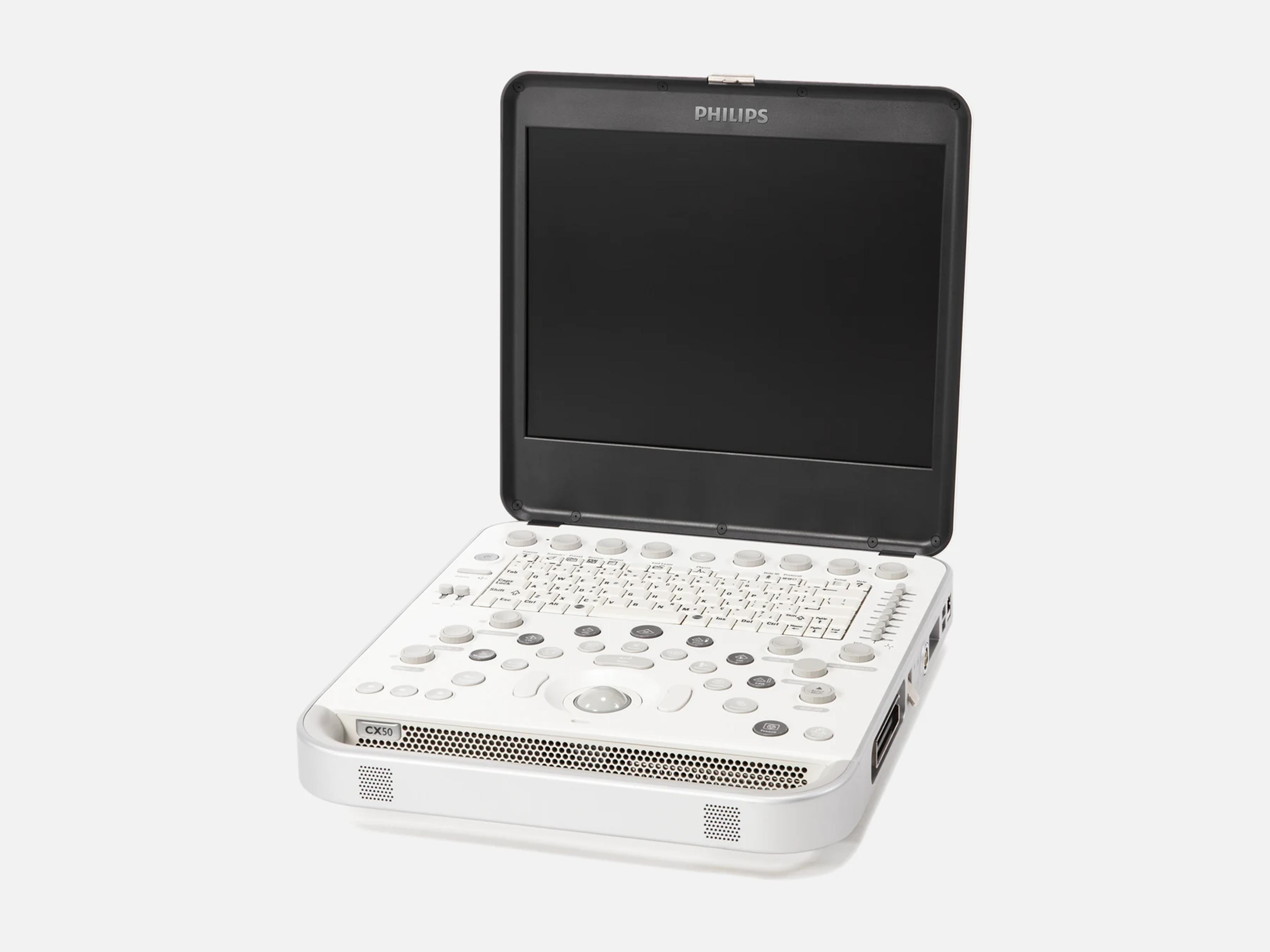
The Philips CX50 is a laptop-style portable ultrasound system carrying technologies from the premium Philips iE33 and iU22 systems. Philips configured it for general imaging, critical care, vascular and regional anesthesia, and the CX50 xMATRIX for diagnostic, interventional, surgical and pediatric echocardiography. Its predecessor is the HD11XE; the cart-based Sparq shares its technology.
- Released
- 2008launched by Philips
- Probe ports
- 1 imaging3 with the multiport adaptor
- Display
- 39.1 cmhigh-resolution LCD
- Weight
- 6.17 kgsystem only
Compatible probes
Linear
- L12-3
- L12-5/50
- L15-7io Compact linear; intraoperative
- L10-4lap Laparoscopic
Convex & microconvex
- C5-1 PureWave
- C8-5
- C9-3io Intraoperative
Phased
- S4-2
- S5-1 PureWave
- S8-3
- S12-4
Endocavity
- C10-3v PureWave; intracavity
Specialty
- X7-2t PureWave TEE; Live 3D on CX50 xMATRIX
- X8-2t PureWave TEE; Live 3D on CX50 xMATRIX
- S7-3t Pediatric and adult 2D TEE
- ViewFlex Xtra Third-party ICE catheter; CX50 xMATRIX
- D2cwc Non-imaging CW probe
- D5cwc Non-imaging static Doppler
Specifications
Display
- Display
- High-resolution LCD, single monitor, 39.1 cm (15.4-inch)
Physical
- Weight
- 6.17 kg, system only; 7.3 kg as specified in 2014
- Weight with cart
- 52 kg, system and cart
- Dimensions
- 41.3 cm wide, 35.6 cm deep, 7.6 cm high (8.6 cm high as specified in 2014)
- Probe ports
1 imaging and 1 pencil-probe port; up to 3 transducers via the multiport adaptorOne imaging-transducer receptacle on the side of the system and a separate pencil-probe receptacle; the optional multiport adaptor on the CX50 cart takes up to three imaging transducers, which attach with multi-pin connectors.Show allShow less
- Controls
Keyboard with Patient and Transducer keys, trackball, Quick Keys, tri-state lightingOne-button controls; keyboard with Patient and Transducer keys; Quick Keys multifunction knobs at the top of the control panel; trackball; tri-state panel lighting showing which controls are available. On CX50 xMATRIX: 3D Zoom/Preview, Live 3D and xPlane rotary controls at the lower left of the panel, and lid-closed operation with tableside control from the Xper Module.Show allShow less
- Carts and carrying
Height-adjustable cart that swivels and locks; travel case; carry handleCX50 cart: system and cart combine into one height-adjustable unit that swivels and locks, with a multiport adaptor for transducers; travel case; hand-carry by the system's handle.Show allShow less
- Control panel height adjustment
- 82.8–107.9 cm
- Packaging weight
- 46.2 kg total: wood 25.5 kg, cardboard/paper 18.4 kg, plastic 2.1 kg, ferro metal 0.2 kg
Power & battery
- Battery
Internal lithium-ion polymer battery with quick-charge; runs on AC or batteryInternal lithium-ion polymer battery with quick-charge battery technology; the system runs on AC or battery, and battery operation allows multiple exams without shutting the system down.Show allShow less
- Power input
- 100–240 V AC, 50/60 Hz, 2.65 A, with the AC adapter
- Power consumption
- 200 W on; 80 W ready to scan or standby; 0 W off
- Energy usage
- 554.8 kWh per year in an average-use scenario
Connectivity & options
- Network
- Ethernet 10/100/1000BaseT and 802.11 a/b/g/n wireless (CX50 5.5)
- DICOM
DICOM to PACS, wired or wireless; storage, worklist, MPPS, print and structured reportsWireless and wired DICOM to PACS; purchasable DICOM services include image storage, storage commitment, structured reports, modality worklist, MPPS and printing; optional ECG and printer peripherals.Show allShow less
- Secure DICOM
- Optional TLS encryption with certificate management (CX50 5.0.X)
- Security
Firewall, OS hardening, malware protection, access controls, patient data encryptionDefense-in-depth security in five layers: firewall, operating system hardening, malware protection, access controls and patient data encryption.Show allShow less
- Export
CD/DVD and USB export with an integrated DICOM viewerCD/DVD and USB study export with an integrated DICOM viewer; on CX50 5.0.X, structured reports cannot be imported.Show allShow less
- Image storage
- Local repository; off-line storage to network, DVR, DVD or USB in JPEG, AVI and DICOM
- Hard drive
- 160 GB, as specified in 2014
- Video output
- DVI-I digital output for cath and EP labs with VGA, DVI-I or HDMI inputs (CX50 xMATRIX)
- Cath lab integration (CX50 xMATRIX)
Xper Module tableside control, EchoNavigator 2.0, automatic patient data exchangeOn CX50 xMATRIX: 2D imaging controlled tableside from the Xper Module of Azurion and Allura Xper systems; EchoNavigator 2.0, an option for Allura and Azurion Xper systems that links Live 3D or Live xPlane images with fluoroscopy; automatic patient data exchange.Show allShow less
- Digital Navigation Link (DNL)
- Real-time image link
- Remote services
ISSL link: proactive monitoring, remote desktop, support request button (optional)ISSL protocol connection to the Philips remote services network over the site's existing Internet access; proactive monitoring, remote desktop support and a control-panel support request button, as optional services that depend on geography and service contract.Show allShow less
- Service tools
- On-cart (non-phantom) transducer test; per-transducer usage reports by exam type
- Remote Reporting
- Qview remote reporting, available only with Qpath (CX50 POC)
- Purchasable imaging options
- LVO Contrast, Physio, Stress Echo Protocol; availability varies by transducer and region
Imaging & software
- Imaging modes
B, M-mode, Anatomical M-mode, PW, CW, Color Doppler, Tissue Doppler, CPA, harmonicsB (2D); M-mode, including Anatomical M-mode; Pulse Wave Doppler; Continuous Wave Doppler; Color Doppler; Color Power Angio (CPA); Tissue Doppler Imaging; tissue harmonic and contrast harmonic imaging. Combined modes B+PWD, B+Color, B+M, B+M+Color, B+Color+PWD, B+CWD and B+Color+CWD. Availability depends on the transducer.Show allShow less
- Revision and software
Cleared releases 1.0, 2.0, 3.0 and 4.0; software labels R1.0.x through v5.5Cleared releases 1.0, 2.0, 3.0 and 4.0; software labels R1.0.x, v1.0.x, R1.1.x, R2.0.x, v2.1.x, R2.5.x, R3.0.x, R4.0.X, 5.0.X and v5.5.Show allShow less
- Beamformer
- Digital broadband beamformer
- Channels
- 128-channel system; 128 transmit and 256 receive channels, 128/128 beamformer
- Digitally processed channels
- Up to 504,576, as specified in 2014
- Dynamic range
- 170 dB full-time system dynamic range
- Image processing
SonoCT compound imaging (up to nine lines of sight), XRES, Advanced XRES, PureWaveSonoCT real-time compound imaging, combining up to nine lines of sight; XRES and Advanced XRES adaptive processing that reduces speckle, haze and clutter; PureWave crystal technology on PureWave transducers.Show allShow less
- Optimization
iSCAN one-button 2D and Doppler (3D on xMATRIX), AutoSCAN, Smart TGC, HD ZoomiSCAN single-button optimization of 2D and Doppler, and of 3D on CX50 xMATRIX; AutoSCAN continuous image-brightness optimization, off by default for 3D TEE on CX50 xMATRIX; Smart TGC predefined time-gain-compensation curves; HD Zoom real-time high-definition zoom.Show allShow less
- Presets
- Tissue-specific presets; FAST and Lung presets
- Active native data
- Scanning parameters adjustable on stored images, clips, 2D and Doppler data
- Doppler tools
- High Q automatic Doppler analysis; Intelligent Doppler holds the selected Doppler angle
- Exam protocols
- SmartExam protocols with on-screen guidance, automatic annotation and report building
- Review
- Cineloop review of buffered images
- CX50 POC tools
Live compare, side-by-side M-mode for lung imaging, Hide patient ID, Temporary IDLive compare, showing live and captured images side by side; side-by-side M-mode with a live reference image for lung imaging interpretation; Hide patient ID for education and presentations; Temporary ID for an interim patient identifier.Show allShow less
- Needle visualization
- On the L12-5 and L12-3 transducers
- Gain Save
- Recalls image settings for each view in stress echo
- 3D
- Freehand 3D volume acquisition
- Live 3D (CX50 xMATRIX)
Live 3D, Live 3D Color, Live 3D Zoom with Preview, Full Volume, Live xPlane, Live 3D TEELive 3D; Live 3D Color; Live 3D Zoom with Preview; Full Volume; Live xPlane, imaging two orthogonal planes at once in grayscale and color Doppler; Live 3D TEE on the X7-2t and X8-2t transducers.Show allShow less
- 3D tools (CX50 xMATRIX)
3D Depth Markers (up to three per volume), 3D Orientation Marker, Auto Crop, Swivel3D Depth Markers, up to three per volume; 3D Orientation Marker; Auto Crop 3D cropping; Swivel display of a frozen 3D image or cine-loop; Target Volume Rate (Target VR) for 3D modes except xPlane.Show allShow less
- Intracardiac echo
- 2D ICE with the ViewFlex Xtra catheter (CX50 xMATRIX)
- QLAB
- Quantification software with Q-Apps
- QLAB cardiac
CMQ, aCMQ A.I. (2D GLS with Auto-ROI EF), MVN A.I., MVQ, Cardiac 3DQ and 3DQA, SQ, TMQCardiac Motion Quantification (CMQ) with speckle tracking; Automated Cardiac Motion Quantification (aCMQ A.I.), 2D Global Longitudinal Strain with Auto-ROI EF, on CX50 xMATRIX; Mitral Valve Navigator A.I. (MVN A.I.), a mitral valve model from a Live 3D volume, on CX50 xMATRIX; Mitral valve quantification (MVQ) from Live 3D TEE; Cardiac 3D Quantification (Cardiac 3DQ) and Cardiac 3D Quantification Advanced (Cardiac 3DQA); Strain Quantification (SQ); Tissue Motion Quantification (TMQ) with speckle tracking.Show allShow less
- QLAB general imaging
IMT, GI 3DQ, MVI, ROI, iSlice (4, 9, 16 or 25 slices)Intima Media Thickness evaluation (IMT); GI 3D Quantification (GI 3DQ); MicroVascular Imaging (MVI); Region of Interest (ROI); iSlice tomographic slicing of acquired volumes in 4, 9, 16 or 25 slices.Show allShow less
Environment
- Heat output
- 700–1100 BTU/hour, fully loaded, as specified in 2014
Compliance
- FDA 510(k)
K201012 (2020-05-01), K162329 (2016-09-14), K123754, K111513, K091804, K081802K201012 (decided 2020-05-01; expanded lung and cardiac imaging labeling for COVID-19 patients); K162329 (2016-09-14; CX50 4.0); K123754 (2012-12-21; CX50 3.0, adding intracardiac echo, intraoperative and laparoscopic indications); K111513 (2011-06-24; pediatric cardiac and neonatal cephalic indications); K091804 (2009-07-17; CX50 2.0); K081802 (2008-07-11; first release). Class II; product codes IYN, IYO and ITX.Show allShow less
- Environmental compliance
- RoHS 2 (EU Directive 2015/863) and REACH
What to check when buying a used Philips CX50
Inspect the power cord and socket for damage
One CX50 power cord, punctured and stripped of its insulation, exposed a live wire that shocked a user lifting the system, and another, pulled too hard, tore its wires out and left the power socket charred.
How: Check the cord end to end for punctures or stripped insulation, and the power socket on the system for charring or loose wires.
Reject a swollen or burned battery
A CX50 battery has been found swollen with its case split, and a burned battery and power supply were traced to a worn-out battery.
How: Take the battery out and look for swelling, a split case or burn marks, and check the system's plastic housing for a seam parting along its weld line.
Inspect every transducer, cable and connector
A transducer or cable showing cracks, splits, fluid leaks, sharp edges or projections must be taken out of use.
How: Examine every part of each transducer, its cable and its connector, and set aside any probe showing that damage.
Check TEE probes against recall Z-1841-2020
Some S7-3t and S8-3t TEE transducers left manufacturing misprogrammed, so an affected probe on a CX50 cannot report its true tip temperature and does not start auto-cool.
How: A CX50 cannot detect an affected probe, so get proof that Philips completed the recall correction on any S7-3t or S8-3t offered with the unit.
Common questions about the Philips CX50
What configurations does the Philips CX50 come in?
Philips offered the CX50 in four clinical configurations: General imaging, Critical care, Vascular and Regional anesthesia. The CX50 POC is the configuration for critical care and emergency medicine, and for regional anesthesia and pain medicine.
The CX50 xMATRIX is the configuration for diagnostic, interventional, surgical and pediatric echocardiography, and it adds Live 3D TEE on the X7-2t transducer. It launched in 2012, and 2D intracardiac echo was added in May 2013.
Is the Philips CX50 discontinued?
Yes, in its four clinical configurations: Philips no longer offers the General imaging, Critical care, Vascular or Regional anesthesia CX50. Philips's Italian catalog still carries the CX50 xMATRIX.
Is the Philips CX50 FDA-cleared?
Yes. The CX50 Diagnostic Ultrasound System is a class II device cleared through FDA 510(k), under product codes IYN, IYO and ITX. Its first release was cleared under K081802 on July 11, 2008.
Later clearances are K091804 for CX50 2.0 (July 17, 2009), K111513 adding pediatric cardiac and neonatal cephalic indications (June 24, 2011), K123754 for CX50 3.0 with intracardiac echo, intraoperative and laparoscopic indications (December 21, 2012), K162329 for CX50 4.0 (September 14, 2016) and K201012, which expanded its labeling for lung and cardiac imaging of COVID-19 patients (May 1, 2020).
What is the difference between the Philips CX50 and the Sparq?
Both use the same technology. The CX50 is built into a portable, laptop-style chassis with an optional cart, while the Sparq is a cart-based mobile system with a capacitive touch interface and an articulating monitor arm. The Sparq has no 3D matrix array transducers.