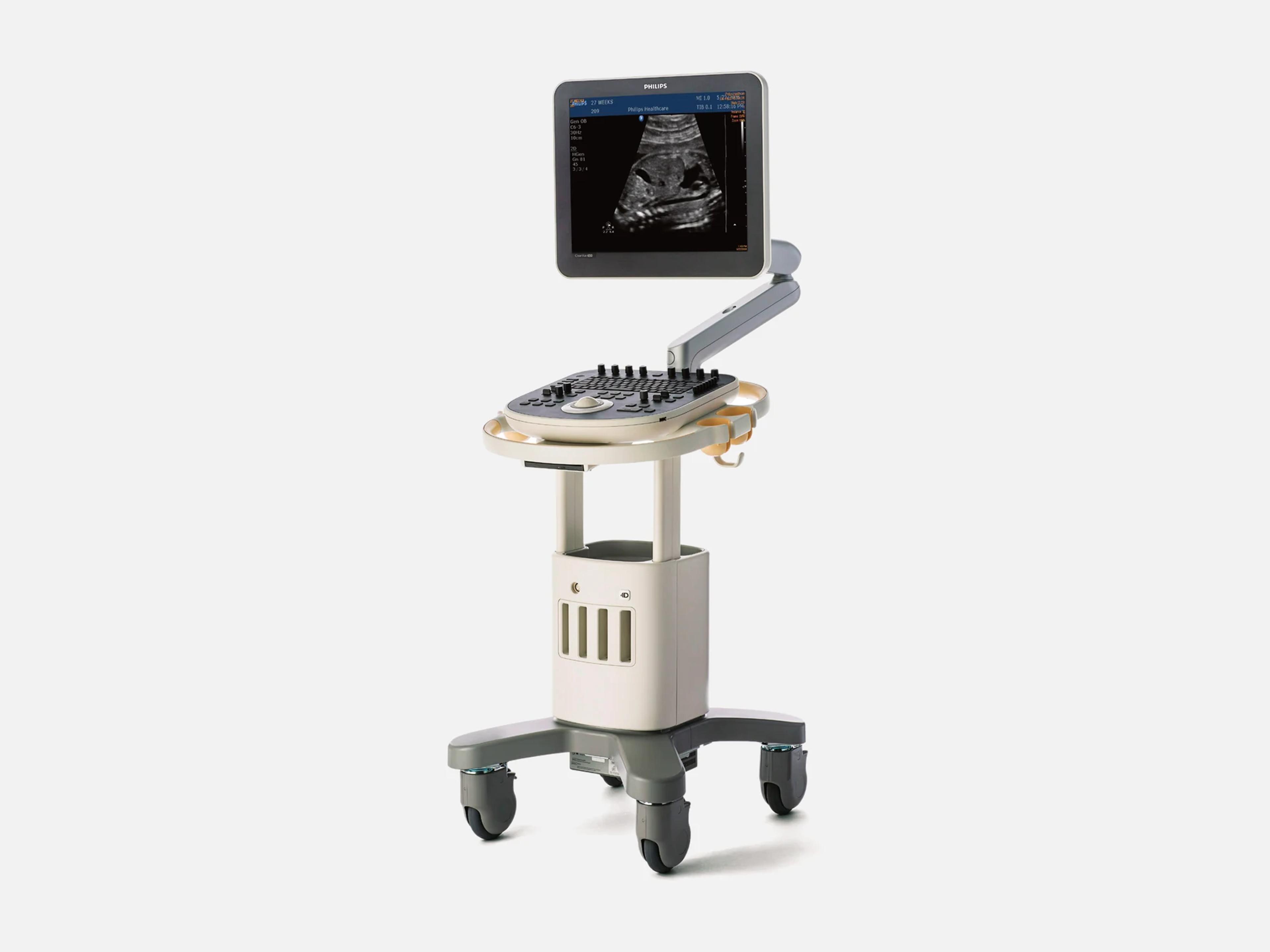
The Philips ClearVue 650 is a cart-based ultrasound system with 2D and 3D/4D imaging for abdominal, Ob/Gyn, vascular, cardiac, small parts, musculoskeletal, urology and pediatric exams. Its ClearVue siblings are the 350 for office use, the 550 for quick scans in busy settings, and the 850 for advanced capability, quantification and ergonomics. It has a 19-inch fold-down LCD and four transducer ports.
- Probe ports
- 4electronically switched
- Display
- 19-inchfold-down LCD, 1280 x 1024
Compatible probes
Linear
- L12-4
- L12-5 38mm Needs software 3.1 or higher
Convex & microconvex
- C5-2
Phased
- S4-1 Sector array
Endocavity
- C9-4v Endovaginal
- 3D9-3v Endovaginal volume array (3D/4D)
Specialty
- V6-2 Volume array (3D/4D)
Specifications
Display
- Display
19-inch fold-down IPS color LCD, 1280 x 1024 at 60 Hz, on a fully articulating arm19-inch (458 x 397 mm) fold-down high-resolution color LCD monitor with an in-plane switching (IPS) panel; SXGA 1280 x 1024 at 60 Hz, non-interlaced RGB; 0.358 mm dot pitch; fully articulating arm with tilt -60°/+90° (fully flat) and swivel +/-90°; backlight lamps contain mercuryShow allShow less
- Brightness
Brightness control with automatic backlight stability (BLS) controlBrightness control with automatic backlight stability (BLS) control, designed for quick warm-up and consistent light output over the display's operational lifeShow allShow less
Physical
- Weight
- 115 lb (52 kg) with printer; total weight 56.43 kg
- Dimensions
W 20.5 in, D 23.0 in, H 45.7 in (arm locked) to 60.7 in (arm raised)Width 20.5 in / 52 cm; depth 23.0 in / 58.4 cm; height 45.7 in / 116.0 cm upright with the arm locked, to 60.7 in / 154.2 cm with the arm fully raised; control panel height 33.0 in / 83.7 cm, not adjustable; the display lifts +6.0 inches from its home positionShow allShow less
- Probe ports
- Four, with electronic switching of up to four imaging transducers; dedicated 4D port
- Controls
QWERTY keyboard, trackball, soft keys, TGC slide pots, LGC soft keys, stereo speakersGraphical user interface; soft keys whose functions change with the active mode, preset or system function; on-screen menus; alphanumeric QWERTY keyboard with globalization key; trackball with Select and Enter keys; imaging mode keys (2D, CPA, M-mode, color, CW, PW, 3D); TGC slide pots and LGC soft keys; integrated stereo speakers; control panel facing the user at a 10° incline, fixed height, with in-context back-lightingShow allShow less
- Cart
Four-wheel swivel, two-wheel lock brake, wrap-around handles, footrest, storage trayFour-wheel swivel; two-wheel lock brake; lightweight cart frame; acquisition module and printer the user can swap; wrap-around handles; integrated footrest; storage tray; cable management hooksShow allShow less
Power & battery
- Power
- 100–240 VAC, 50–60 Hz; 450 VA power, 300 VA consumed; built-in A/C line conditioner
- Battery
- Button cell on the motherboard
Connectivity & options
- Input/output ports
3 x USB, composite video, S-video, VGA, LAN (RJ-45), EKG plugThree USB ports (optional footswitch, data transfer, qualified plain paper printers); black and white composite video output; S-video output; VGA output; LAN connector (RJ-45) for DICOM networking and Philips Remote Services; EKG plugShow allShow less
- DICOM
Option: DICOM print and store, worklist, MPPS, structured reporting, storage commitmentDICOM networking option over the LAN connector: DICOM 3.0 print and store service class user, multiple DICOM servers and presets, modality worklist, modality performed procedure step (MPPS), DICOM structured reporting for vascular, cardiac and Ob/Gyn; storage commitment (push model) at software 1.0; at release 3.1, ultrasound image and multiframe storage, storage commitment, Comprehensive SR, modality worklist, MPPS and grayscale and color print management are each a purchasable optionShow allShow less
- Data storage
320 GB hard drive; DVD/CD write and read; export to USB; cineloop up to 1,200 frames320 GB hard drive; DVD/CD write and read (multi-session, supports 4.7 GB DVD); single frames and loops stored to the internal hard disk, USB flash and CD/DVD; cineloop of up to 1,200 frames of 2D and color imagesShow allShow less
- Philips Remote Services
Remote clinical and technical support; needs a service agreement and internet accessPhilips Remote Services connectivity for virtual on-site clinical and technical support (service agreement and internet access required); optional online support request, optional proactive monitoring and optional Utilization Reports; Remote Desktop where availableShow allShow less
- Printer
- Digital B/W thermal printer (USB input); plain paper printers supported
- Footswitch
- Optional three-pedal USB footswitch: freeze, acquire, print
- Barcode scanner
- Optional; enters patient data from hospital-generated barcodes
- ECG
- One three-lead ECG input; ECG-triggered skipping between 1 and 20 beats
Imaging & software
- Imaging modes
2D, CPA, color, M-mode, Anatomical M, Color M, PW, HPRF PW, TDI, CW, 3D/4D, STIC2D (Microfine 2D focusing); Color Power Angio (CPA) and Directional CPA; color Doppler; M-mode; Anatomical M-mode; Color M-mode; pulsed wave Doppler; High PRF pulsed wave Doppler; Tissue Doppler Imaging (TDI); continuous wave Doppler (cardiac sector transducers only); Freehand 3D; automated 3D/4D imaging; Fetal STIC; color compare mode; dual mode; duplex; triplex (2D, Doppler, and color or CPA); Tissue Harmonic Imaging (THI); Pulse Inversion Harmonic (PIH); panoramic; trapezoidalShow allShow less
- Software level
Software 1.0, 3.0 and 3.1; a 3.3 level is listed for the ClearVue 350/550/650/850Software releases 1.0, 3.0 and 3.1, with release 3.1 shared across the ClearVue 350, 550, 650 and 850; a 3.3 level is also named for all four modelsShow allShow less
- Applications
Abdominal, Ob/Gyn, vascular, cardiac, small parts, MSK, urology, pediatric, TCDAbdominal, Ob/Gyn, vascular, cardiac, small parts, musculoskeletal, urology, pediatric and TCDShow allShow less
- System architecture
All-digital broadband beamformer; 32,768 processed channels; 170 dB input dynamic rangeAll-digital broadband beamformer; Microfine 2D focusing with dynamic focal tuning; 170 dB full-time input dynamic range; 32,768 digitally-processed channels; 256 gray shades (8 bits); acquisition frame rate greater than 1128 frames per second in high frame rate mode, dependent on transducer, field of view, depth and angleShow allShow less
- Maximum imaging depth
- To 30 cm, exam and transducer specific
- Advanced 3D/4D imaging with MPR
Option: automated 3D, 4D and MPR on volume transducers; Freehand 3D on all transducersOption: automated 3D, 4D and multiplanar reformatting (MPR) imaging on volume transducers; quantitative 3D volume acquisition on the V6-2 and 3D9-3v; Freehand 3D on all imaging transducersShow allShow less
- 3D fetal echo STIC
Option on volume transducers; grayscale only or combined with color Doppler3D fetal echo STIC (Spatio-Temporal Image Correlation) for fetal echocardiography, an option on volume transducers; image capture in grayscale only or combined with color DopplerShow allShow less
- Auto Face Reveal
Option: removes tissue over the fetal skull to show the face with one buttonOption: a segmentation tool that detects a sphere representing the fetal skull and trims the overlying tissue (3D trim) to show the fetal face with one buttonShow allShow less
- SonoCT
Real-time compound imaging on the C5-2, C9-4v, L12-4, V6-2 and 3D9-3vReal-time compound imaging with beam-steered compounding (up to nine lines of sight), an option together with XRES; available on the C5-2, C9-4v, L12-4, V6-2 and 3D9-3vShow allShow less
- XRES
Adaptive image processing; 2D, CPA, M-mode, dual, CW Doppler, zoom, post-Freeze, loopsXRES adaptive image processing, an option together with SonoCT; available in 2D, Color Power Angio, M-mode, dual imaging, CW Doppler, zoom, post-Freeze and when capturing loopsShow allShow less
- Tissue Harmonic Imaging with Pulse Inversion
- THI on S4-1, C5-2, V6-2, L12-4; Pulse Inversion Harmonic (PIH) on C5-2, S4-1, V6-2
- 2D Opt
2X multi-line parallel processing, frequency compounding; key with up to three settings2D Opt signal processing with 2X multi-line parallel processing and frequency compounding; a 2D Opt key with up to three settings for patient-specific optimization in 2D and color DopplerShow allShow less
- Advanced system automation
iSCAN, 2D Opt, High Q, Intelligent Doppler, Adaptive Doppler, Adaptive color DopplerOption package: 2D, color and PW Doppler iSCAN, 2D Opt, High Q, Intelligent Doppler, Adaptive Doppler and Adaptive color DopplerShow allShow less
- iSCAN
Automatic TGC and gain in 2D, gain in color, one-button spectral optimization in PWiSCAN intelligent optimization: in 2D, automatic adjustment of TGC and receiver gain; in PW Doppler, one-button optimization of spectral tracing; in color, automatic adjustment of receiver gainShow allShow less
- High Q automatic Doppler analysis
Automatic real-time and retrospective tracing of peak and mean velocityHigh Q automatic Doppler analysis: automatic real-time and retrospective tracing of instantaneous peak velocity and intensity-weighted mean velocityShow allShow less
- Intelligent Doppler
Maintains angle-to-flow automatically; linear transducers, vascular and general imagingAutomatically maintains angle-to-flow for Doppler velocity measurements; available with the vascular and general imaging application packages, on linear transducers onlyShow allShow less
- Anatomical M-mode
- Option on all sector transducers; M-mode trace from a user-defined line
- Chroma imaging
- Multiple color maps for 2D and M-mode colorization
- Panoramic and 3D imaging
- Option: extended field of view and freehand 3D surface rendering
- Trapezoidal imaging
- Up to 21° on each side on linear arrays, in vascular and general imaging
- Active Array technology
- Core feature, available on all ClearVue transducers, including the V6-2 and 3D9-3v
- SmartExam protocols
Option: guided protocols for adult echo, abdominal, GYN and lower extremity; customizableSmartExam system-guided protocols, an option: an on-screen exam guide with the required views by exam type; preset protocols for adult echo, abdominal, GYN and lower extremity; fully customizable protocolsShow allShow less
- Clinical analysis packages
General imaging, Ob/Gyn and fertility, cardiac and vascular analysis, with reportsAnalysis packages for general imaging, Ob/Gyn and fertility, and cardiac and vascular, with application-specific reports; OB workflow tools with quick-launch OB measurements and configurable reports and analysisShow allShow less
- On-cart maintenance tools
Software maintenance that keeps presets and patient data; transducer assurance testingOn-cart software maintenance tools that optimize, maintain and repair system software while preserving annotations, calculations, presets and patient data; on-cart transducer assurance testingShow allShow less
Environment
- Operating environment
10–40 °C, 15–80% RH non-condensing; heat dissipation <700 BTU/hour fully loadedSystem and printer: 10–40 °C at 15–80% relative humidity (non-condensing); heat dissipation <700 BTU/hour (fully loaded)Show allShow less
Compliance
- FDA 510(k)
K153480 (decided 2015-12-16); K120321 (ClearVue 350/550 family)K153480, decided 2015-12-16, issued under the ClearVue 850 name, with the ClearVue 350, 550, 650 and 850 listed under it; K120321, the ClearVue 350/550 clearance, which a later FDA filing extends to the 650. Class II; product codes IYO (regulation 892.1560) and IYN (regulation 892.1550)Show allShow less
- Electrical safety standards
- AAMI/ANSI ES60601-1; IEC 60601-1; CSA C22.2 No. 60601.1; EN 60601-1
Languages
- Languages
English, French, German, Italian, Japanese, Portuguese, Russian, Spanish…English, French, German, Italian, Japanese, Portuguese, Russian, Spanish and Simplified ChineseShow allShow less
What to check when buying a used Philips ClearVue 650
Test the monitor arm and its mounting screws
An FDA report on this model records a monitor that detached from its articulation arm, and service found a loose connection at the arm's mounting screws.
How: Move the monitor through its full tilt and swivel, and look for play where it meets the arm, loose or missing mounting screws, and oil around the arm's pressurized gas strut.
Inspect the screen for cracks and uneven light
The monitor's backlight lamps contain mercury, so a damaged monitor has to be handled and disposed of with care.
How: Check the whole screen for cracks, then raise the brightness and look for dim patches or uneven light across the panel.
Inspect each probe's cable and strain relief
Wiping a probe's cable or strain relief with isopropyl alcohol can damage it, and Philips does not cover that damage under its warranty or a service contract.
How: Look along each cable and at its strain reliefs for cracks, splits or discoloration, and ask which disinfectants were used and whether each is approved for that transducer.
Check the hard disk for prior patients' exams
Patient data, presets, annotations and calculations are kept on the system itself, so a used unit can arrive holding the previous owner's patient records.
How: Review the exams saved on the internal hard disk, and settle before purchase who removes the prior patients' data and which presets stay.
Common questions about the Philips ClearVue 650
Is the Philips ClearVue 650 FDA-cleared?
Yes. The ClearVue 650 is listed with the FDA as a class II diagnostic ultrasound system under 510(k) K153480, decided on December 16, 2015, together with the ClearVue 350, 550 and 850; that clearance was issued under the ClearVue 850 name. A later FDA filing also places the 650 under K120321, the clearance first issued for the ClearVue 350/550. Its product codes are IYO and IYN.
Does the Philips ClearVue 650 have 3D/4D imaging?
Yes, though not necessarily on every unit. The ClearVue 650 is positioned as a system with 3D/4D from the start, but advanced 3D/4D imaging with multiplanar reformatting (MPR) is an option on it.
Automated 3D, 4D and MPR imaging run on the volume transducers, the V6-2 and the 3D9-3v, while Freehand 3D works on every imaging transducer. On a used unit, ask to see 4D acquisition working with the volume probe that comes with it.
Does the Philips ClearVue 650 include DICOM?
As an option: DICOM networking on the ClearVue 650 runs over its LAN connector. At software release 3.1, ultrasound image and multiframe storage, storage commitment, Comprehensive SR, modality worklist, MPPS and grayscale and color print management are each a purchasable option, so ask which of them a used unit has enabled and see each one work on your network.
Is the Philips ClearVue 650 discontinued?
Yes. The ClearVue 650 came out in 2013, when it joined the ClearVue 350 and 550 as the newest model in the family, and it has since been discontinued.