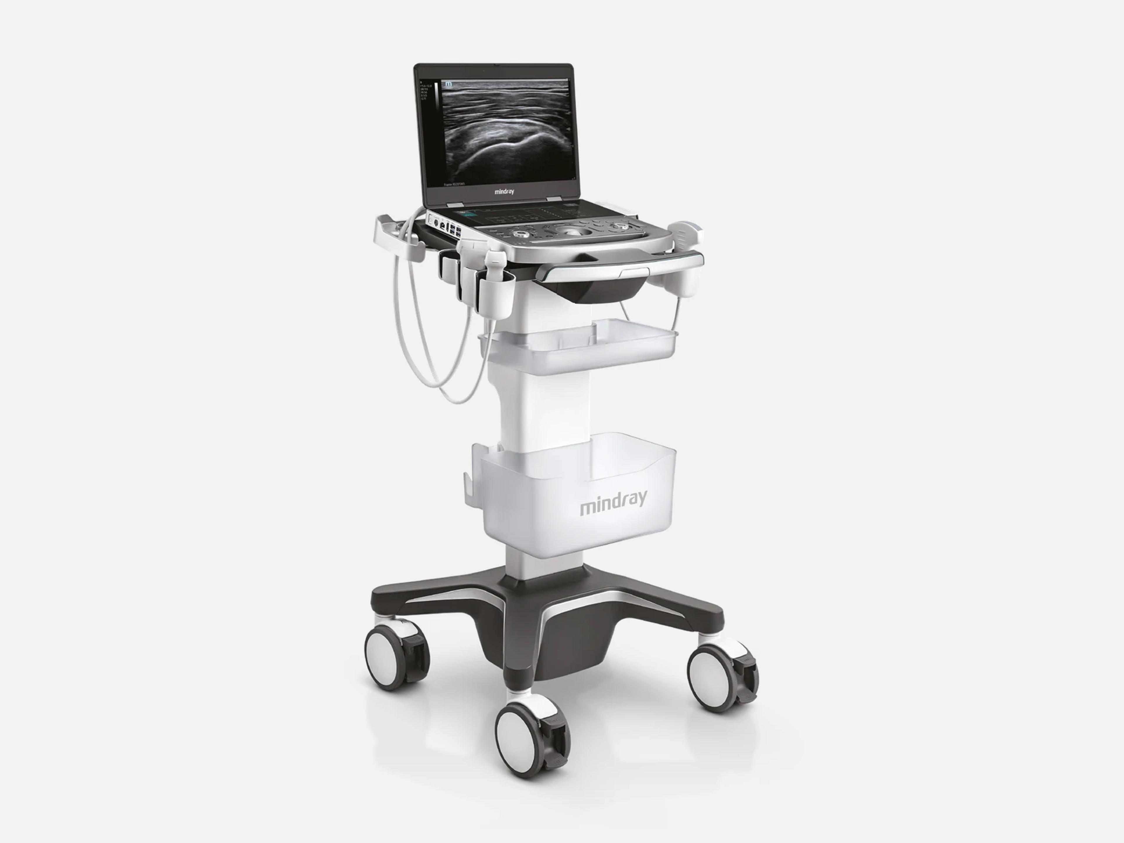
The Mindray MX8 is a portable laptop ultrasound system for cardiovascular or musculoskeletal imaging. Its MSK configuration has needle tools such as iNeedle+ and eSpacial Navi™, and its Cardiovascular configuration has Auto EF, tissue tracking and LVO with Stress Echocardiography. It weighs 6.6 lb, has a 15.6-inch LED monitor and shares FDA 510(k) clearances with siblings including the MX7, MX7T, ME7 and ME8.
- Released
- 2020first FDA clearance
- Display
- 15.6-inch1920 × 1080 LED monitor
- Weight
- 6.6 lbunder 4.0 kg with batteries
Compatible probes
Linear
- L9-3s Cardiovascular and MSK configurations
- L11-3VNs MSK configuration; works with eSpacial Navi™
- L12-3RCs Three programmable buttons; Cardiovascular and MSK
- L13-3Ns MSK configuration
- L14-3Ws MSK configuration
- L14-6Ns MSK configuration
- L15-3RCs Cardiovascular and MSK configurations
- L16-4Hs MSK configuration
- L20-5s MSK configuration
- L24-6Hs MSK configuration
- L24-6RCs Cardiovascular and MSK configurations
Convex & microconvex
- SC5-1Ns Cardiovascular and MSK configurations
- SC10-2RCs MSK configuration
Phased
- P8-2s Cardiovascular configuration
- P10-4s Cardiovascular configuration
- SP5-1Ns Cardiovascular configuration
Specialty
- P7-3Ts Transesophageal (TEE); Cardiovascular configuration
- CW2 Non-imaging CW pencil probe; Cardiovascular configuration
Specifications
Display
- Display
15.6-inch LED monitor, 1920 × 1080; fold-down LCD; secondary status display15.6-inch LED monitor with 1920 × 1080 resolution; fold-down LCD display, and folding it fully puts the system into standby after 30 seconds; secondary display showing battery capacity, AC power status and product modelShow allShow less
- Touch screen
- 12.3-inch LED touchscreen, 1920 × 720, customizable
Physical
- Weight
- 6.6 lb; under 4.0 kg including batteries
- Dimensions
- (322±5) × (364±5) × (44±3) mm (D × W × H); 1.7 in thick
- Probe ports
One main-unit port with a locking lever; the Dual Probe Extend Module expands it to twoA probe port on the main unit with a probe locking lever, accepting a probe or a probe extend module; the Dual Probe Extend Module expands it to two connections, and the optional Probe Adapter PCM-CWS01 connects the CW pencil probe to the portShow allShow less
- Controls
- Trackball or trackpad, touch screen, user-defined keys, iVocal voice control
Power & battery
- Battery
Built-in lithium-ion, 14.4 V, 6600 mAh; 90 minutes of continuous B-mode scanningBuilt-in lithium-ion battery: 90 minutes of continuous scan time in B-mode; 14.4 V, 6600 mAh (single battery); charging from 0 to 100% takes less than 4 hours with the system powered off or in standbyShow allShow less
- U-Bank battery
Optional; up to 4 hours of continuous scanning, with 2 or 4 batteries; 19 V DCOptional U-Bank battery companion: up to 4 hours of continuous scanning; 2 or 4 batteries; 19 V DC; 26400 mAh or 13200 mAh; charging from 0 to 100% takes about 5 hours with the system powered off or in standbyShow allShow less
- Power supply
- External adapter, 100–240 V AC, 50/60 Hz; input 2.0–1.0 A, 4.4–2.5 A on the cart
Connectivity & options
- DICOM
DICOM 3.0: worklist, MPPS, storage commitment, query/retrieve, print, structured reportsDICOM 3.0: verification; storage (US, multi-frame, secondary capture, encapsulated PDF, raw data, enhanced US volume); modality worklist; MPPS; storage commitment; query/retrieve; basic color and grayscale print; comprehensive SR storage, with structured reports for OB-GYN, echocardiography, vascular, breast and abdomen; HL7 query V2.3 to V2.6Show allShow less
- Network
- Single network interface, Ethernet 10/100/1000BaseT; optional WiFi module
- Ports
S-Video out, network, HDMI, USB ports, power inputS-Video output, network port, HDMI, USB ports (for storage, a bar code reader, a printer, a footswitch and a DVD recorder) and a power input portShow allShow less
- Storage
Internal hard disk; SSD storage; DICOM media storage to CD-R, DVD and USB devicesInternal hard disk for images and patient data; SSD storage; DICOM media storage to CD-R, DVD and USB devicesShow allShow less
- iStorage
- Needs external UltraAssist software version 2.0 with the V1.0 network protocol
- Standard configuration
Main unit, power adapter, built-in batteries, system software, traveling caseMain unit with a carrying handle, power adapter, built-in batteries, system software, traveling case and accessories (operator's manuals, user-defined label, cables)Show allShow less
- Hardware options
Mobile cart, ECG module, WiFi module, Probe Adapter PCM-CWS01, U-Bank, Air Capsule…Mobile cart (with power supply and retractable cable, with power supply without retractable cable, or without power supply); ECG module with ECG cables; WiFi module; Probe Adapter PCM-CWS01 (with CW2s); U-Bank (2 or 4 batteries); barcode reader; Air Capsule (with wireless probes); iVocal Microphone; iVocal Plus Microphone Array; Magnetizer package (for eSpacial Navi); footswitch; multilingual controls overlay; dust-proof, main unit and display coversShow allShow less
- Peripherals
Video printers, iVocal headsets and mics, DVD R/W drive, barcode readers, footswitchesVideo printers P95DW-N, UP-X898MD and UP-D25MD; iVocal headsets and microphones XPD1 Headset, XPD1 Presentation and PUSBMIC43; external DVD R/W drive GP60NB60; barcode readers DS4308, LS2208, HS-1M, HS-1R and HH1800; footswitches 971-SWNOM (2 or 3 pedals) and FS-81-SP-2 (1 pedal)Show allShow less
Imaging & software
- Imaging modes
B, M, PWD, CWD, Color, Power, TDI, Smart 3D, 4D, Strain Elastography, contrast, iScape…B, M, PWD, CWD, Color Doppler, Amplitude (Power) Doppler, combined modes (B+M, PW+B, Color+B, Power+B, PW+Color+B, Power+PW+B), Tissue Harmonic Imaging, iScape, TDI, color M, Smart 3D, 4D (real-time 3D), Strain Elastography and contrast imaging (LVO and liver contrast agents)Show allShow less
- Imaging technology
- ZONE Sonography® Technology+ (ZST+)
- HD Scope (Cardiovascular configuration)
- Enhances B-mode tissue characterization in a selected region of interest
- Auto EF (Cardiovascular configuration)
Recognizes diastole and systole frames in 2D echo data and calculates EDV, ESV and EFAnalyzes 2D echo data to recognize diastole and systole frames and calculate EDV, ESV and EF for left ventricle function evaluationShow allShow less
- Tissue Tracking with Quantitative Analysis (TTQA) (Cardiovascular configuration)
Tracks speckle frame to frame to analyze cardiac tissue motion, not angle-dependentAnalyzes cardiac tissue motion by tracking speckle displacement frame to frame; not angle-dependent, with onboard analysisShow allShow less
- LVO with Stress Echocardiography (Cardiovascular configuration)
- Customizable user protocols, wall motion scoring and a reporting suite
- RIMT (Cardiovascular configuration)
- Automatic carotid intima-media thickness measurement from real-time radio frequency data
- iNeedle+ (MSK configuration)
Detects needle angle and improves needle visibility on linear and convex transducersSecond-generation needle visualization: automatically detects needle angle and improves needle visibility on linear and convex transducers during interventional proceduresShow allShow less
- eSpacial Navi™ (MSK configuration)
4D magnetic needle navigation for in-plane or out-of-plane procedures, with the L11-3VNs4D magnetic needle navigation for in-plane or out-of-plane procedures; compatible with the L11-3VNs transducer, and supported by the Magnetizer package optionShow allShow less
- iVocal voice control (MSK configuration)
Second-generation AI voice recognition for hands-free scanningSecond-generation iVocal voice control based on AI voice recognition, for hands-free scanning; the iVocal Microphone and the iVocal Plus Microphone Array are optionsShow allShow less
- iWorks
Standardized exam protocols, including MSK protocols; user-created examsiWorks Protocols: pre-labeled, standardized exam protocols, including MSK protocols for wrist, shoulder, hip, knee, elbow and ankle; user-created exams with 2D, M-mode, Doppler, measurements and annotationsShow allShow less
- Living Technology™
- Software-based upgrades; new application packages and hardware are optional purchases
- Operating system and data security
- Windows 10 with Windows Defender; hard-disk drive encryption and secure data wipe
- Options and application packages (family list)
iScape View, Free Xros M, Free Xros CM, Tissue Doppler Imaging, Contrast imaging, LVO…For the family that shares the MX8's operator's manuals, not split by model or configuration: iScape View, Free Xros M, Free Xros CM, Tissue Doppler Imaging, TDI QA, CW, Contrast imaging, Contrast Imaging QA, LVO, Low MI Contrast, Strain Elastography, Stress Echo, Tissue Tracking QA, Smart 3D, 4D, iLive, TTQA-RV, Smart Face, DICOM Basic and DICOM options; Abdomen/General, Obstetrics, Gynecology, Cardiology, Small Parts, Urology, Vascular, Pediatrics, Nerve and Emergency&Critical packages; RIMT, AutoEF, R-VQS, Smart B-line, Smart VTI, Smart IVC, CPP, Smart FHR OB1, Smart Bladder, Auto DFR, Auto GA, Auto VM, Smart FV, HD Access Works, VA Grid, iWorks, iNeedle, eSpacial Navi, iVocal and DVR ModuleShow allShow less
Environment
- Operating
- 0 °C to 40 °C; 20% to 85% relative humidity, no condensation; 700 hPa to 1060 hPa
- Storage and transport
- -20 °C to 55 °C; 20% to 95% relative humidity, no condensation; 700 hPa to 1060 hPa
Compliance
- FDA 510(k)
K251192 (2025-08-22), K241432 (2024-09-12), K212900 (2021-11-05), K200001 (2020-04-09)K251192 (decided 2025-08-22); K241432 (2024-09-12); K212900 (2021-11-05); K200001 (2020-04-09). Each includes the MX8; K212900 and K241432 clear it under a device name shared with sibling models such as the MX7, MX7T, ME7 and ME8. Class II, 21 CFR 892.1550, product codes IYN, IYO and ITX.Show allShow less
- Safety classification
Class I, internally powered; Type-BF applied part; main unit IPX0; power adapter IP22Class I equipment and internally powered equipment; Type-BF applied part; main unit IPX0; i3P, i5M and i8L probes IP68, other probes IPX7; power adapter IP22; portable equipment, and mobile equipment when installed on the mobile cart; continuous operationShow allShow less
What to check when buying a used Mindray MX8
Have a biomed run electrical safety tests
Long use can degrade electrical and mechanical performance, with leakage, distortion and abrasion, and reduce image sensitivity and precision.
How: With a safety analyzer, measure protective earth resistance and earth, enclosure, patient and mains-on-applied-part leakage.
Inspect each probe's head, lens and cable
A defective probe can give the patient an electric shock, and a probe with a damaged or scratched surface or exposed wiring must not be used.
How: Look for cracks or swelling on the head, peeling, protruding parts or a loose acoustic lens, solidified gel on the lens, and deterioration, cracks or peeling on the cable and connector.
Check each probe for abnormal heat while scanning
A probe that gives off excessive heat can burn the patient.
How: Scan with every probe offered and feel its surface for abnormal heat.
Look for residue of spilled fluid
The system is not waterproof, and fluid that gets into the monitor or system can damage it and cause electric shock or system failure.
How: Check the monitor and the system casing for any residue left by a fluid spill.
Common questions about the Mindray MX8
Is the Mindray MX8 FDA-cleared?
Yes. The MX8 is FDA 510(k)-cleared as a Class II device under 21 CFR 892.1550, with product codes IYN, IYO and ITX.
Clearances that include it are K200001 (April 9, 2020), K212900 (November 5, 2021), K241432 (September 12, 2024) and K251192 (August 22, 2025). K212900 and K241432 clear it together with sibling models, among them the MX7, MX7T, ME7 and ME8.
What is the difference between the Mindray MX8 MSK and Cardiovascular configurations?
The MSK Ultrasound System is designed with musculoskeletal imaging in mind and features needle visualization tools: iNeedle+ detects the needle angle and improves needle visibility on linear and convex transducers, and eSpacial Navi™ adds 4D magnetic needle navigation with the L11-3VNs transducer.
The MX8 Cardiovascular Ultrasound System is designed with cardiovascular imaging in mind, with cardiac tools such as Auto EF, TTQA tissue tracking and LVO with Stress Echocardiography, and RIMT for carotid intima-media thickness. Each configuration has its own transducer family; the Cardiovascular one includes the P7-3Ts transesophageal probe and the CW2 non-imaging CW pencil probe.
How is the Mindray MX8 different from the ME8?
The two share FDA 510(k) clearances and operator's manuals. The MX8 is a specialty system for cardiovascular or musculoskeletal imaging, while the ME8 was introduced as a point-of-care ultrasound system.
What warranty does the Mindray MX8 come with?
Bought directly from Mindray North America, the MX8 carries a standard five-year warranty on the system, its parts and standard transducers, while specialty transducers, TEE probes included, carry one year. Terms for a system bought from one of Mindray's agents can vary.
The warranty does not cover parts or accessories Mindray has not approved, repairs by anyone outside Mindray's service network, or an instrument or part whose serial number is not legible enough.