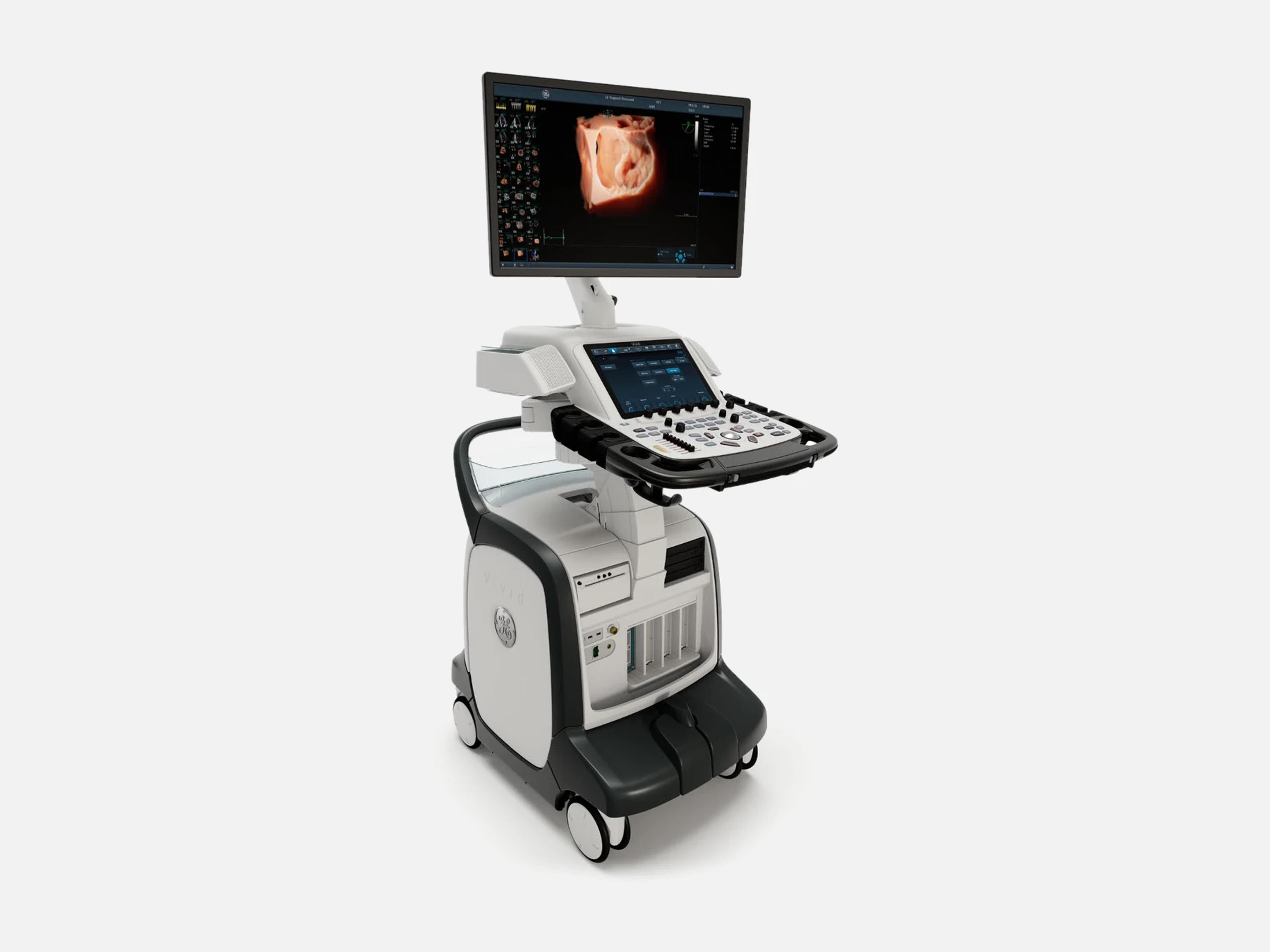
The GE Vivid E95 is a premium 4D cardiovascular ultrasound system, specialized for cardiac imaging, with vascular and general radiology applications. It has GE's cSound software beamformer and five probe ports, one for CW pencil probes, and runs 4D transthoracic and TEE probes. Its siblings, the Vivid E90 and E80, need an option for 4D imaging on v206 software; the 4V-D probe is listed for the E95 alone.
- Released
- 2015first FDA clearance
- Probe ports
- 54 imaging, 1 CW Doppler
- Display
- 23.8-inch HDUUltra Edition configuration
- Weight
- 120 kg ± 10%264 lb, with monitor only
Compatible probes
Linear
- 9L-D Vascular
- 11L-D Vascular
- ML6-15-D
- L8-18i-D
Convex & microconvex
- C1-6-D XDclear; abdominal, renal, urology, OB/GYN
- C2-9-D XDclear; abdominal, renal, urology, OB/GYN
- C3-10-D
- 8C
Phased
- 4Vc-D 4D transthoracic; needs software v202 r.46.0 or higher
- 4V-D 4D; listed for Vivid E95 only
- M5Sc-D
- 6Vc-D Pediatric 4D transthoracic; needs software v204 or higher
- 6S-D Pediatric
- 12S-D Pediatric
Endocavity
- iC5-9-D
Specialty
- 6VT-D 4D transesophageal, adult interventional
- 9VT-D Mini 4D transesophageal, pediatric and interventional; introduced with v206
- 10T-D Neonatal micro multiplane transesophageal
- 6Tc Transesophageal; uses the DP port
- 6Tc-RS Transesophageal
- 9T Transesophageal
- 9T-RS Transesophageal
- P2D CW Doppler pencil; uses the Doppler port
- P6D CW Doppler pencil; uses the Doppler port
- 4D ICE NUVISION 4D intracardiac catheter; US only
Specifications
Display
- Main monitor
19-inch LCD, 22-inch OLED, 24-inch LCD or 23.8-inch HDU, depending on configuration19-inch LCD, 22-inch widescreen OLED (MCM-2250NB), 24-inch LCD (introduced for Com Express with software v203) or 23.8-inch HDU (High Definition Ultrasound) on an articulating monitor arm in the Ultra Edition configuration. Units delivered with v206 have an LCD or OLED main monitor; the HDU spare is specified only for systems originally shipped with HDU.Show allShow less
- Touch panel
- 12-inch high-resolution LCD multi-touch screen with adjustment rotaries
- Monitor arm
- Swivels, folds into a locked position, and adjusts fore/aft and vertically
- External monitor
- From v203 (Com Express) hardware, the OS detects it and software duplicates the screen
Physical
- Weight
120 kg ± 10% (264 lb ± 10%) with monitor, without probes and peripherals120 kg ± 10% (264 lb ± 10%) with monitor, without probes and peripherals (v206); 128 kg (283 lb) or more when ready for use, depending on installed peripherals.Show allShow less
- Dimensions
Transport position: H 141.0 cm (55.5 in), W 58.5 cm (23 in), D 83.0 cm (32.7 in)In transport position, with monitor and peripherals: height 141.0 cm (55.5 in), width 58.5 cm (23 in), depth 83.0 cm (32.7 in).Show allShow less
- Probe ports
Five: four imaging-probe connectors and one Doppler port for CW pencil probesFive probe ports: one DP port (for the 6Tc TEE probe with a Vivid 7 connector), three DLP ports (Vivid E95/E90/E80 and Vivid E9 probe connectors) and one Doppler port for CW pencil probes. The four imaging connectors comprise one 128-channel connector, two 192-channel connectors and one that takes 192- or 256-channel probes.Show allShow less
- Top Console
Motor-driven lift; moves side to side and back and forth; Park Lock for transportThe Top Console (operating panel, monitor and keyboard) moves up and down (motor-driven, gas-spring assisted), side to side and back and forth, with motor-operated X/Y brakes and a Park Lock for transport; single-hand push for up/down and left-right adjustment.Show allShow less
- Mobility
Four swivel casters; front pedal brake and direction lock; individual rear brakesFour swivel casters; a pedal mechanism at the front operates the parking brake and the direction lock on the front wheels; the rear casters have individually operated brakes; grab handles front and rear.Show allShow less
- Operating panel
TGC sliders, trackball, scan-mode, measure and store keys; keys lit by availabilityOn/off button, touch panel, TGC sliders, trackball area, scan-mode, measurement and image-store keys and loudspeaker volume; keys are lit by availability. Electronically extendable, and height- and side-adjustable.Show allShow less
- Keyboard
- QWERTY keyboard in a drawer below the operating panel; adjustable floating keyboard
- Trackball
- Optical
- Footswitch
- 3-button
- Speakers
- Stereo sound during Doppler scanning and replay
Power & battery
- Power
100–240 VAC ±10%, 50/60 Hz; 700 W with monitor and on-board peripherals100–240 VAC ±10%, 50/60 Hz; power consumption 700 W including monitor and on-board peripherals; up to 3 A at 230 VAC and up to 7 A at 100 VAC; needs a dedicated single branch circuit.Show allShow less
- Backup battery
- Lithium-ion, on some configurations
Connectivity & options
- Network
Wired Ethernet (optional 10/100/1000-Mbit DICOM); wireless LAN through a GE USB kitWired Ethernet on the rear I/O panel, with optional 10/100/1000-Mbit Ethernet/DICOM networking; wireless LAN through a GE USB adapter kit.Show allShow less
- DICOM
Storage, Modality Worklist, Query/Retrieve, Structured Reporting and printingDICOM Storage, Modality Worklist, Query/Retrieve and Structured Reporting (measurements), plus DICOM printing; the system exchanges data with EchoPAC, Image Vault and other DICOM servers.Show allShow less
- Patient inputs
- ECG, PHONO, AUX (pressure/pulse); ECG synchronizes images and is not a patient monitor
- Printers
- Internal UP-D898DC; external UP-D25MD and Color LaserJet Pro M454dn
- Video and media
Optional internal DVR; external HVO-550MD DVR; DVD multi drive (DV-W5600S-400)Optional internal digital video recorder; external HVO-550MD digital video stream recorder; DVD multi drive (DV-W5600S-400) connected by an eSATA-USB-C cable.Show allShow less
- Remote service
- InSite remote connection to GE online service and applications engineers
- Software delivery
- Through GE's service platform or an end-user portal, with entitlement
- Streaming
StreamServer option (sector and TEE probes only); Remote Viewing, not for diagnosisStreamServer option key streams live 2D and 3D ultrasound data over the network, with sector and TEE probes only; Remote Viewing streams the main monitor over the local network and is not for diagnostic use.Show allShow less
- Cloud
- Tricefy Uplink for archiving and sharing (registration required); Tricefy QR for review
- Fleet data
- Imaging Insights: a dashboard of system and operator usage built from DICOM data
- Security
Disk encryption (customer choice), whitelisting (always on), LDAP, SonoDefenseDisk encryption, enabled at the customer's choice; application whitelisting, enabled at the factory and impossible to change or disable; LDAP authentication, where streaming access needs a group mapping to ConsultingPhys; SonoDefense.Show allShow less
- Training
- Digital Expert education and training support, separate from the console
Imaging & software
- Revision and software
Versions v201, v202, v203, v204 and v206; revisions written as r-numbers (v202 r.46.0)Software versions 201, 202, 203, 204 and 206; system and application software on v206 read v206.xx, and revisions are written as r-numbers, for example v202 r.46.0.Show allShow less
- Operating system
- Windows 10 Embedded (v206)
- Beamformer
cSound software beamformer; cSound ADAPT corrects speed-of-sound aberrations (4Vc-D)cSound software beamformer; cSound 2.0 updates arrived in 2017/18 and cSound 3.0 in 2019. cSound ADAPT continuously optimizes beamforming to correct aberrations from speed-of-sound differences; it was launched for the 4Vc-D probe and is exclusive to the Vivid E95 and Vivid E90.Show allShow less
- Processing
192 channels transmitted, received and processed simultaneously; two GPU cards192 channels, transmitted, received and processed simultaneously. Back End Processor with two GPU cards: GPU-1 for beamforming and GPU-2 for display acceleration; systems delivered with v206 use an A4000 and a T600.Show allShow less
- Imaging modes
2D, M, Color M, Color/Power Doppler, PW/CW, TVI, strain, TSI, contrast, 4D, multi-plane2D (B) mode, M-mode, Color M-mode, Color Doppler, Power Doppler (Angio), PW and CW Doppler, Tissue Velocity Imaging (TVI), Tissue Tracking, strain rate, strain, Tissue Synchronization Imaging (TSI), contrast, 4D and multi-plane modes; harmonic imaging, coded pulse and combined modes (B/M, B/Color M, B/PWD or CWD, B/Color/PWD or CWD, B/Power/PWD).Show allShow less
- Maximum imaging depth
- Up to 50 cm, depending on probe type and clinical application
- Maximum frame rate
- 6656 frames per second
- Cine memory
- Up to 500 MB
- Internal storage
- 2.5-inch SATA hard disk; 1 TB 2.5-inch SATA SSD as a v206 renewal part
- 4D acquisition
- Single Beat 4D; Vmax single-beat 4D volume rates, enabled by cSound 2.0
- 4D quantification
4D Auto LVQ, 4D Auto RVQ, 4D Auto LAQ, 4D Auto MVQ, 4D Auto TVQ, 4D Auto AVQ4D Auto LVQ (left ventricle), 4D Auto RVQ (right ventricle), 4D Auto LAQ (left atrial volumes and ejection fraction), 4D Auto MVQ (mitral valve and annulus, TEE or TTE), 4D Auto TVQ (tricuspid valve), 4D Auto AVQ (aortic outflow tract); Advanced 4D User Quantification Package; Advanced 4D User Toolbox, which includes FlexiSlice.Show allShow less
- Strain and ejection fraction
AFI 3.0, AFI LA, AFI RV, Auto EF 3.0, Easy AutoEF, Easy AFI LV, MyoCardial WorkAFI 3.0 (2D speckle-tracking LV function, including 2D Auto EF, at rest or with AFI stress, on GE raw data and DICOM images from other systems), AFI LA, AFI RV, Triplane AFI, Auto EF 3.0, Easy AutoEF (adult TTE on GE raw B-mode LV loops; not for LVs with septal bulge), Easy AFI LV (AI-based global and segmental LV strain with view recognition), AI Auto ROI, MyoCardial Work (global longitudinal strain considered with arterial blood pressure); AFI 2.0 and AutoEF 2.0; optional 2D Auto EF 3.0 and AFI 3.0 Productivity Package.Show allShow less
- Stress echo
- 2D Stress, 4D Stress, Multiplane Stress, Smart Stress, AFI Stress
- Automation and AI
AI Auto Measure – 2D, AI Auto Measure – Spectrum Recognition, Cardiac Auto Doppler, ASOAI Auto Measure – 2D (semi-automatic LV measurements from the parasternal long axis), AI Auto Measure – Spectrum Recognition (a Doppler trace and measurements in two clicks), Cardiac Auto Doppler, Automatic View Recognition, Automatic Spectrum Optimization (ASO, which sets PW/CW baseline and scale when Auto is pressed), Auto Tissue (Soft and Sharp), Dynamic Auto Optimization, Scan Assist Pro, QuickApps (instant recall of stored settings, keeping the live image geometry), Smart Depth, Continuous Tissue Optimization (CTO).Show allShow less
- Flow imaging
Blood Speckle Imaging 2.0, HD Color, BFI, B-Flow, Real-time 3D Color FlowBlood Speckle Imaging (BSI) and Blood Speckle Imaging 2.0 (flow visualization that reduces angle dependence and aliasing), HD Color (4D color Doppler with transparency control for low-flow components), Blood Flow Imaging (BFI), B-Flow, Real-time 3D Color Flow, Pulsatile-Flow.Show allShow less
- Rendering and navigation
FlexiLight, HDlive, FlexiViews, FlexiSlice Extend, Live FlexiSlice 2.0, Dual CropFlexiLight (light-source photo-realistic rendering of heart structures, live and in playback), HDlive (light and shadow effects in tissue), FlexiViews, FlexiSlice, FlexiSlice Extend (navigation for structural heart procedures), Live FlexiSlice 2.0 (three 2D planes from the 4D volume, live during the exam), Dual Crop (structures from two directions at once), Slice View, StereoVision, Laser-Lines.Show allShow less
- Interventional tools
4D ICE, 4D Markers, Pre-Post Compare, CT Fusion Live, 2-click crop, Biplane prepare4D ICE (a compatible catheter is required and market restrictions apply), 4D Markers (markers for communication between interventionalist and echo imager), Pre-Post Compare (pre- and post-procedure images and measurements), CT Fusion and CT Fusion Live (live co-registered navigation in 4D ultrasound and CT data), 2-click crop, 4D views, Biplane prepare, View-X (an external video signal such as fluoroscopy, streamed over the network into a window on the screen, without audio).Show allShow less
- Image processing
UD Clarity, UD Speckle reduce, Hybrid Filter, TCI, Virtual Apex, Virtual ConvexUD Clarity (cardiac speckle reduction), UD Speckle reduce (non-cardiac), Hybrid Filter, True Confocal Imaging (TCI: automatic focusing through the field of view, highlighted with the 9L-D and 11L-D linear probes), Virtual Apex (probe-dependent near-field imaging), Virtual Convex (linear probes), spatial compounding and speckle reduction imaging, SI/SRI, Extended FOV, LOGIQView panoramic imaging, Advanced Contrast Low MI, Adaptive Contrast Enhancement, Strain Elastography.Show allShow less
- Measurement and display tools
Zscore, Spline Tool, DICOM PDF Read, Image View, LaunchpadZscore value sets; Spline Tool (area measurement); DICOM PDF Read (read-only access to DICOM PDF reports on a DICOM server); Image View (duplicates the main-monitor image on the touch screen); Launchpad (shows and launches qualified third-party apps installed on the system).Show allShow less
- Probe Check
- Tests each probe's element integrity in air; it does not catch every defect
Environment
- Operating environment
- 10–35 °C (50–95 °F); 30–85% humidity, non-condensing; 700–1060 hPa
- Storage and transport
- −20 to +60 °C (−4 to +140 °F); 10–95% humidity, non-condensing; 700–1060 hPa
- Heat output
- Up to 2390 BTU/h with monitor and on-board peripherals
- Cooling
- Low-noise cooling; fan speed follows internal temperature; air filter on rear intake
- Acoustic noise
- 29 dB (minimum) to 42 dB (maximum)
Compliance
- FDA 510(k)
K150087 (2015-03-04), K170823, K173341, K181685, K200743, K202658, K220882 (2022-07-22)K150087 (decided 2015-03-04); K170823 (2017-06-12); K173341 (2017-11-02); K181685 (clearance letter 2018-10-25); K200743 (2020-07-23); K202658 (2021-03-05); K220882 (2022-07-22). Class II; product codes IYN, IYO and ITX; 21 CFR 892.1550; first predicate the Vivid E9 (K131514).Show allShow less
- Cleared applications
Cardiac, vascular, fetal/OB, abdominal, pediatric, small organ, MSK, urology, TEE…Fetal/obstetrics; abdominal (including renal and GYN); pediatric; small organ (breast, testes, thyroid); neonatal and adult cephalic; cardiac (adult and pediatric); peripheral vascular; musculoskeletal, conventional and superficial; urology (including prostate); transesophageal; transvaginal; transrectal; intracardiac and intra-luminal; interventional guidance (including biopsy and vascular access); thoracic/pleural; intraoperative (vascular).Show allShow less
What to check when buying a used GE Vivid E95
Inspect the power cord and its system inlet
Recall Z-2988-2018 corrected Vivid E95 systems whose power cord may break at the system end and expose its conductors.
How: Confirm the recall correction was completed on the unit offered, then check the cord and connectors for damage, correct line, neutral and ground wiring, and a working inlet retainer.
Have a biomed run GE's electrical safety tests
Electrical leakage from the console and from each probe has acceptance limits drawn from the medical electrical safety standards.
How: Test the system and every probe offered for leakage, and compare the results with the unit's earlier safety-test records.
Confirm both DICOM Worklist recall corrections were applied
Recalls Z-1779-2017 and Z-2316-2024 concern the wrong patient being shown or selected when the DICOM Worklist responds slowly.
How: Check whether the unit's serial number was covered and corrected under each recall, and before scanning confirm the selected patient's details match the patient.
Reject a damaged, leaking or deformed backup battery
Some E95 configurations carry a lithium-ion backup battery, so find out whether the unit offered has one.
How: Where one is fitted, look for damage, leakage, deformation, discoloration or an odor.
Common questions about the GE Vivid E95
Is the GE Vivid E95 FDA-cleared?
Yes. The Vivid E95 was first FDA-cleared under 510(k) K150087, decided March 4, 2015, as a class II device (product codes IYN, IYO and ITX; 21 CFR 892.1550). Later clearances shared with the Vivid E90 and Vivid E80 include K200743 (July 23, 2020), K202658 (March 5, 2021) and K220882 (July 22, 2022).
How does the Vivid E95 differ from the Vivid E90 and E80?
On v206 software, the Vivid E90 and Vivid E80 offer 4D imaging only with the 4D option; the Vivid E95 carries no such condition. The 4V-D probe is listed for the Vivid E95 only, while cSound ADAPT and the 9VT-D mini 4D TEE probe are limited to the E95 and E90. CT Fusion Live is exclusive to the three systems.
Can the GE Vivid E95 be used with an ICE catheter?
Yes. The 4D ICE NUVISION intracardiac catheter is listed for the Vivid E95 and Vivid S70N, with availability restricted to the USA. 510(k) K202658, decided March 5, 2021, added intracardiac and intra-luminal applications and the catheter as a compatible accessory, with a tool that recognizes it.
Has the GE Vivid E95 been recalled?
Three FDA recalls apply to the Vivid E95. Z-1779-2017 concerned DICOM Worklist patient selection and was terminated in 2018; Z-2988-2018 concerned the system-side power cord and was terminated in 2021; Z-2316-2024, posted in July 2024, concerns DICOM Worklist response time.