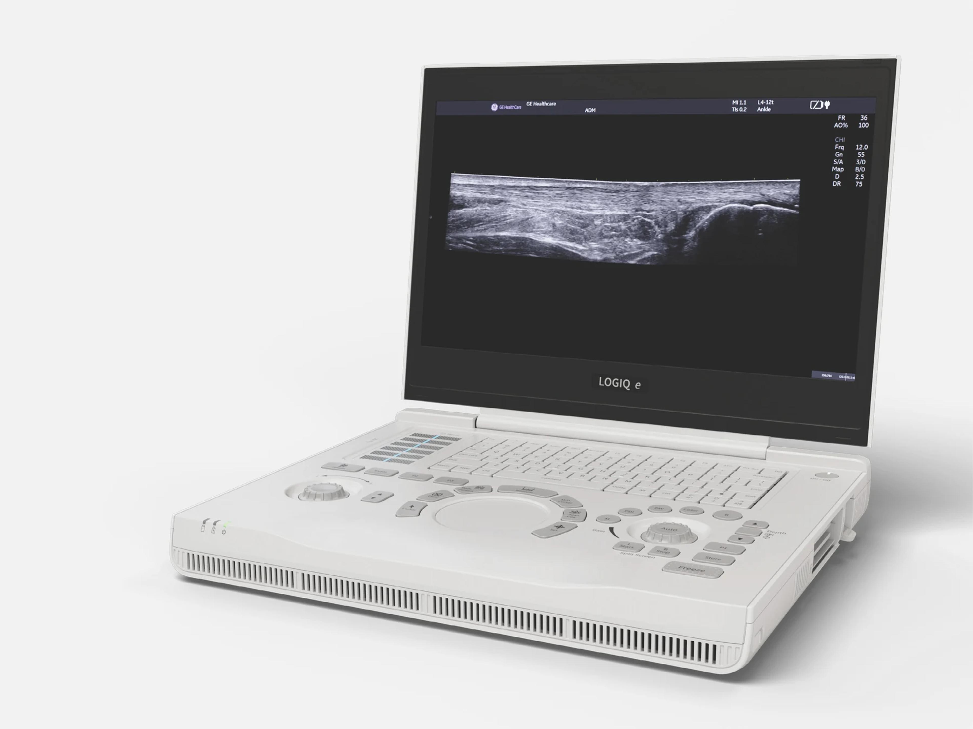
The GE LOGIQ e R9 is a laptop-style portable ultrasound system that weighs 4.5 kg with its battery and has a 15.6-inch color display. GE designs the LOGIQ e as a general-purpose system for hospitals and clinics, with features for anesthesiology, musculoskeletal, rheumatology, endocrinology and vascular labs. It runs R11.x.x software and shares its service manual with the LOGIQ Xe and LOGIQ He systems.
- Probe ports
- 1 on consoleup to 3 with cart option
- Display
- 15.6-inchcolor LCD, 1920x1080
- Weight
- 4.5 kg9.92 lb, with battery
Compatible probes
Linear
- 9L-RS · 3–9 MHz
- 12L-RS · 4.2–13 MHz
- L4-12t-RS · 4.2–13 MHz Four configurable buttons
- ML6-15-RS · 5–15 MHz Active matrix
- L4-20t-RS · 5–20 MHz XDclear, four configurable buttons
- L8-18i-RS · 6.7–18 MHz Hockey stick
- L10-22-RS · 10–22 MHz
Convex & microconvex
- C1-5-RS · 2–5 MHz
- 4C-RS
- 8C-RS · 4.2–11 MHz Microconvex
Phased
- 3Sc-RS · 1.7–4 MHz
- M5Sc-RS
- 6S-RS · 3–7 MHz
- 12S-RS · 4.1–12 MHz
Endocavity
- E8C-RS · 4.2–10 MHz Transrectal, transvaginal
Specialty
- 6Tc-RS · 3–8 MHz Transesophageal
Specifications
Display
- Display
- 15.6-inch color LCD, 1920x1080 pixels, ±85° horizontal and vertical viewing angle
- Display modes
Simultaneous B/PW, B/CFM or PDI, B/M, Dual B, real-time Triplex; split and quad screenSimultaneous: B/PW or TVI/TVD, B/CFM or PDI, B/M or AMM, Dual B (B/B), Dual B + CFM or PDI, Needle Recognition + CFM (PDI), real-time Triplex. Also multi-image split screen (live and/or frozen), quad-screen display, timeline display, colorized B/M/PW/CW.Show allShow less
- Display arrangements
Split screen, independent cine playback, conventional/widescreen display, read/write zoomSplit-screen live/frozen views, independent cine playback, conventional/widescreen display and read/write zoomShow allShow less
Physical
- Weight
4.5 kg (9.92 lb) console with battery; 5.2 kg or more ready for useConsole with battery: 4.5 kg (9.92 lb), or 4.5 ± 0.2 kg (9.92 ± 0.44 lb). Ready for use: 5.2 kg or more, depending on installed peripherals.Show allShow less
- Dimensions (console only)
- Height 47 mm (1.85 in), width 295 mm (11.61 in), length 375 mm (14.76 in)
- Cart dimensions
- Height 840–1140 mm (33.0–44.9 in), width 510.3 mm (20.1 in), depth 510.3 mm (20.1 in)
- Probe ports
1 on the console, with locking lever; up to 3 with the optional 3-probe port on the cartConsole: one probe connector port with a locking lever. Up to three probe ports with the optional 3-probe port on the LOGIQ e docking cart.Show allShow less
- Controls
Sealed alphanumeric keyboard, backlit keys, touch-panel TGC, trackpad; trackball optionSealed alphanumeric keyboard with ergonomic, backlit hard keys; digital TGC on an illuminated, waterproof touch panel; trackpad (scrolling, measurement and analysis, position/size, scan-area width, tilt, multi-touch gain and depth), with a trackball option; user-defined function keys and programmable image store keys; user-programmable presets; brightness and audio volume controls; interactive dynamic software menu.Show allShow less
Power & battery
- Power (console)
- 100–240 VAC, 50/60 Hz, max 150 VA, through an external AC/DC adapter
- Battery
- Lithium-ion; two internal battery packs in the console
- Battery charge time
Full charge in 210 minutes; 5% to 80% in 5 hours or less while live scanningConsole batteries: full charge in 210 minutes. Both internal packs: 5% to 80% in 5 hours or less while the system is live scanning.Show allShow less
- Standby
- A fully charged battery lasts 180 days in standby
- Extended battery module (cart option)
3 hours scanning; 4.5 hours with the console batteries; full charge in 3.5 hoursOptional extended battery module on the cart: 3 hours scanning when fully charged; 4.5 hours scanning together with the console batteries; full charge in 3.5 hours; 180 days in standby.Show allShow less
- Boot-up time
- 25 seconds from standby
Connectivity & options
- Console connections
- 3 × USB 3.0 (max 5.0 Gbps), LAN 10/100/1000 BaseT, HDMI 1.4, DC power input
- Docking cart connections
- 4 × USB 2.0, AC power input
- Wireless
- Wireless LAN through an optional USB wireless adapter
- DICOM
- DICOM 3.0 connectivity, encrypted DICOM, DICOM TLS
- Security
Patient-data encryption, DICOM TLS, whitelisting, LDAP, account policies, access controlEncryption for patient data; DICOM TLS; whitelisting; LDAP interface for user credentials; customized policies for user accounts; security for access control.Show allShow less
- Hardware options
Footswitch, printer, USB ECG (AHA/IEC), barcode scanner, wireless adapter, DVD R/WFootswitch; printer; USB ECG (AHA/IEC); barcode scanner; wireless adapter; external DVD R/W storage.Show allShow less
- Cart options
- 3-probe port, extended battery, printer bracket, storage basket
- Approved peripherals
Printers, DVD-RW, 3-pedal footswitch, USB wireless adapter, ECG-USB, barcode scannerUP-D898MD B/W printer; UP-D25MD color printer; TS8XDVDS-K DVD-RW; MKF 2-MED GP26 3-pedal footswitch; A8000 USB wireless adapter; ECGUSB1D-EX ECG-USB; XENON 1950 barcode scanner.Show allShow less
- Remote service
- InSite ExC capability
- eDelivery
Software updates over GE's service platform (Insite RSvP) or by portal downloadOn-device software update delivery through GE's service platform (requires Insite RSvP connectivity), or by download from an end-user portal and local installation.Show allShow less
- Imaging Insights
Utilization and operator-usage dashboard from DICOM data of GE and other systemsCollects DICOM data from GE and other makers' ultrasound equipment and shows system utilization and operator usage on a dashboard.Show allShow less
Imaging & software
- Revision and software
- R9; R11.x.x software, on the R11.0.x branch (R11.0.4 and above)
- Computer platform
- Duo core processor; Windows 10 operating system
- Clinical applications
Abdominal, fetal/OB, pediatric, small organ, cephalic, cardiac, peripheral vascular, MSKOphthalmic; fetal/OB; abdominal (gyn and urology); pediatric; small organ (breast, testes, thyroid); neonatal and adult cephalic; cardiac (adult and pediatric); peripheral vascular; musculoskeletal conventional and superficial; transrectal; transvaginal; transesophageal; intraoperative (abdominal, thoracic, peripheral); thoracic/pleural for motion and fluid detection; imaging guidance of interventional procedures (e.g. nerve block, vascular access).Show allShow less
- Imaging modes
B, M, AMM/Color M, CFM, PDI, PW, CW, M-CFM, 3D; options: High-Res PDI, TVI/TVDB-Mode; M-Mode; Anatomical M-Mode (AMM)/Color M-Mode; Color Flow Mode (CFM, Color Doppler); Power Doppler Imaging (PDI); Pulsed Wave Doppler (PW); Continuous Wave Doppler (CW); M-Color Flow Mode (M-CFM); 3D; optional High-Res PDI and Tissue Velocity Imaging/Doppler (TVI/TVD).Show allShow less
- B-Mode
Depth 0.5–33 cm; gain 0–90 dB; dynamic range 36–96 dB; up to 8 focal zones; 12 gray mapsDepth 0.5–33 cm (probe-dependent); gain 0–90 dB in 1 dB steps; dynamic range 36–96 dB in 3–6 dB increments; up to 8 focal zones (probe-dependent); 12 gray-scale maps; maximum frame rate ≥1598 Hz.Show allShow less
- Doppler
PW max 870 cm/s; CW max 1,460 cm/s; angle correct ±90°; CFM PRF 0.3K–22K HzPW: max 870 cm/s (19,800 Hz), min 15 cm/s. CW: max 1,460 cm/s (40,000 Hz), min 40 cm/s. Angle correct ±90° in 1° steps. CFM PRF 0.3K–22K Hz (probe-dependent); CFM maximum frame rate 330 Hz.Show allShow less
- Contrast
- Optional operating mode; not available in the U.S.
- Coded Harmonic Imaging (CHI)
Enhances near-field resolution and far-field penetration; reduces low-frequency noiseTissue harmonic imaging: raises near-field resolution for small parts and far-field penetration, and reduces low-frequency amplitude noise.Show allShow less
- CrossXBeam
Spatial compounding, 3, 5, 7 or 9 angles; live side-by-side DualViewSpatial compounding with 3, 5, 7 or 9 angles; live side-by-side DualView display; works with Color Doppler, PW, SRI-HD, Coded Harmonic Imaging and Virtual Convex.Show allShow less
- Speckle Reduction Imaging (SRI-HD)
- Multiple speckle-reduction levels; works with B-Mode, Color Doppler and 3D
- Virtual convex and virtual apex
- Wider far-field view: virtual convex on linear probes, virtual apex on sector probes
- LOGIQ View
- Panoramic image up to 60 cm in long axis from a manual sweep
- 3D
- B-Mode and CFM rendered in 3D from acquired color data; 3D landscape and 3D movie
- Anatomical M-Mode (AMM)
- M-Mode on stored 2D cine clips
- M-Color Flow Mode (M-CFM)
- Color overlaid on the M-Mode trace
- Tissue Velocity Imaging/Doppler (TVI/TVD) (option)
- TVI color-codes tissue velocities; TVD gives spectral data for a Doppler sample
- High-Res PDI (option)
Power Doppler mode combining B-Mode and Color Flow Doppler effectsOptional power Doppler mode combining B-Mode and Color Flow Doppler effects for hemodynamics visualization; available on the 9L-RS, 12L-RS, L4-12t-RS, L8-18i-RS, L10-22-RS, ML6-15-RS and L4-20t-RS probes.Show allShow less
- Needle recognition (option)
Adjustable needle gain and angle, in Color and Power Doppler; on linear and convex probesOptional. Displays the needle, anatomy and motion, including in Color and Power Doppler; needle gain and angle adjustable; available on all linear and convex probes.Show allShow less
- Strain elastography
- Elastography designed to assist treatment planning and monitoring
- Automatic optimization
ATO (tissue), ACO (color flow), ASO (spectrum)Auto Tissue Optimization (ATO), Auto Color Flow Optimization (ACO) and Auto Spectrum Optimization (ASO).Show allShow less
- Patient follow-up tool with fusion (option)
Recalls the previous exam's parameters, comments, body patterns; warns of a probe changeOptional. Recalls the previous exam's imaging parameters, comments and body patterns, and alerts if a different transducer is used; works in B-Mode, Color Mode and PDI.Show allShow less
- Calculations
Cardiac, gynecological, OB, multi-gestational, MSK, hip dysplasia, pediatric, renalCardiac, gynecological, multi-gestational, musculoskeletal and hip dysplasia, OB (with tables), pediatric, renal, rheumatology, small parts, urological and vascular calculations.Show allShow less
- Measurement and analysis
- Full M&A calculation package with real-time auto Doppler calculations
- Auto IMT (option)
- Automated intima-media thickness of the common carotid artery
- Auto EF (option)
- Automated ejection fraction measurement
- Flow quantitative analysis (option)
- Quantifies blood flow within a region of interest
- Software options
Auto EF, Auto IMT, easy3D, eSmart trainer, Stress echo, Ophthalmic, Scan assistantAuto EF, Auto IMT, easy3D, eSmart trainer, Flow quantitative analysis, High-res PDI, LOGIQ View, Needle recognition, Ophthalmic, Patient follow-up tool with fusion, Scan assistant, Strain elastography, Stress echo, TVI/TVD.Show allShow less
- eSmart Trainer (option)
- Training modules: scanning technique, probe-position graphics, anatomy, example images
- Standard features
Loop storage, on-board help, patient database, Probe Check, quick save, report writerLoop storage from live scanning and from memory; on-board user manual (Help); patient information database; probe check; quick save; raw data processing; report writer package; scan assistant; Speckle Reduction Imaging (SRI-HD); strain elastography; virtual convex/virtual apex.Show allShow less
- Probe Check
- Tests each probe element's response while scanning in air
- Quick save
- One button sends an image or a whole exam to a memory stick or the network
- Report writer
On-board reports: exam data, measurements, calculations, images; customizable templatesOn-board reporting: patient and exam information, measurements, calculations, images and comments in a report for printing or review on a standard PC; standard templates, customizable.Show allShow less
- Presets
- User-programmable presets; factory default preset data
- Cine memory
- 256 MB
- Storage
- Integrated solid-state drive
- Other features
- Fine angle steer, HD zoom, customizable user interface, fetal trending
- Probe loading
- Less than 5 seconds
Environment
- Operating (with probe)
- 10–40 °C (50–104 °F), 30–75% humidity non-condensing, 700–1060 hPa
- Storage temperature
- -5 to 50 °C
- Transport temperature
- -20 to 50 °C
Compliance
- FDA 510(k)
K232186 (2023-11-03); earlier K151028, K133533, K113690, K072797K232186 (LOGIQ e, decided 2023-11-03); K151028 (LOGIQ e, 2015-06-18); K133533 (LOGIQ E, 2014-02-12); K113690 (LOGIQ i, LOGIQ E and Vivid e, 2011-12-29); K072797 (LOGIQ E compact ultrasound, with LOGIQ i and Vivid e, 2007-10-17).Show allShow less
- Safety and standards
AAMI/ANSI ES60601-1, CAN/CSA-C22.2 No. 60601-1; CE marked; IEC 60601-1, -1-2, -1-6, -2-37Certified to AAMI/ANSI ES60601-1:2005/(R)2012 and CAN/CSA-C22.2 No. 60601-1:14; CE marked to Council Directive 93/42/EEC; conforms to IEC 60601-1, IEC 60601-1-2, IEC 60601-1-6, IEC 60601-2-37, ISO 10993 and NEMA UD3 acoustic output display.Show allShow less
Languages
- User interface languages
English, Chinese, French, German, Japanese, Russian, Spanish and eight moreBrazilian Portuguese, Chinese, Danish, Dutch, English, Finnish, French, German, Greek, Italian, Japanese, Norwegian, Russian, Spanish, Swedish.Show allShow less
What to check when buying a used GE LOGIQ e R9
Inspect the power cord and AC/DC adapter
An adverse-event report on a LOGIQ e adapter describes heat and arcing at the power-cord connector, attributed to foreign conductive material in the connection.
How: Look over the cord, plug, appliance inlet and adapter for damaged insulation, weak strain relief, debris or heat marks, and confirm the adapter's label voltage matches your site.
Have a biomed run the electrical safety tests
Ground continuity and leakage tests show whether the console and each probe are safe to use on patients.
How: Ask for ground continuity from the plug's ground pin to exposed metal, chassis leakage and each probe's leakage current, and if a probe fails, retest it on another connector or swap probes to tell a probe fault from a console fault.
Reject a damaged, leaking or deformed battery
The console carries internal lithium-ion battery packs, and a damaged or leaking pack is a safety fault rather than wear.
How: Inspect each battery pack for damage, leakage, an odor, deformation or discoloration.
Test boot, battery operation and the logs
A unit that will not run on battery points to a broken battery, a poor battery connection or a charger-board fault, and the logs show faults a short demonstration can miss.
How: Boot it from AC and from battery, waiting after each shutdown before powering on again, then run it on battery alone with probes disconnected until it shuts down, recharge it, and review the error, temperature and configuration logs and any battery-expiry reminder on screen.
Common questions about the GE LOGIQ e R9
Is the GE LOGIQ e R9 FDA-cleared?
The LOGIQ e is FDA-cleared under 510(k) K232186, decided on November 3, 2023.
Earlier 510(k) clearances naming the LOGIQ e are K151028 (June 18, 2015), K133533 (February 12, 2014), K113690 (December 29, 2011) and K072797 (October 17, 2007); the last two also name the LOGIQ i and Vivid e. The clearances name the LOGIQ e, not a particular revision.
Does the GE LOGIQ e R9 offer contrast imaging in the US?
No. Contrast is an optional operating mode of the LOGIQ e, and it is not available in the United States.
What are the GE LOGIQ e R9 console model numbers?
GE designates the LOGIQ e consoles H48942CC (TB Basic Console), H48942CD (TP Basic Console), H48942CH (TP CKD console), H48322DB (TP CKD with Probe Check) and H48322DC (TP Basic with Probe Check).
The LOGIQ e Vet Basic Consoles are H48942CE (TB) and H48942CF (TP).
Which GE systems share the LOGIQ e R9's service manual?
The LOGIQ e basic service manual for R11.x.x software also applies to the LOGIQ Xe Expert, Pro and Performance; the LOGIQ He Expert, Pro, Performance, Elite, Super, Standard, Surgery and Intervention; and the LOGIQ e Exp, LOGIQ e Pro and LOGIQ e Vet.